Knee
Learn about the most common knee conditions that we can help you treat
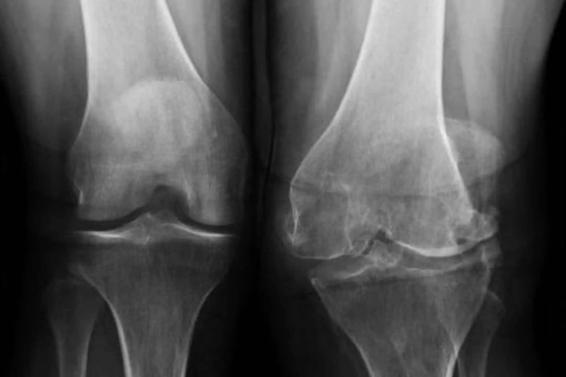
Osteoarthritis of the Knee
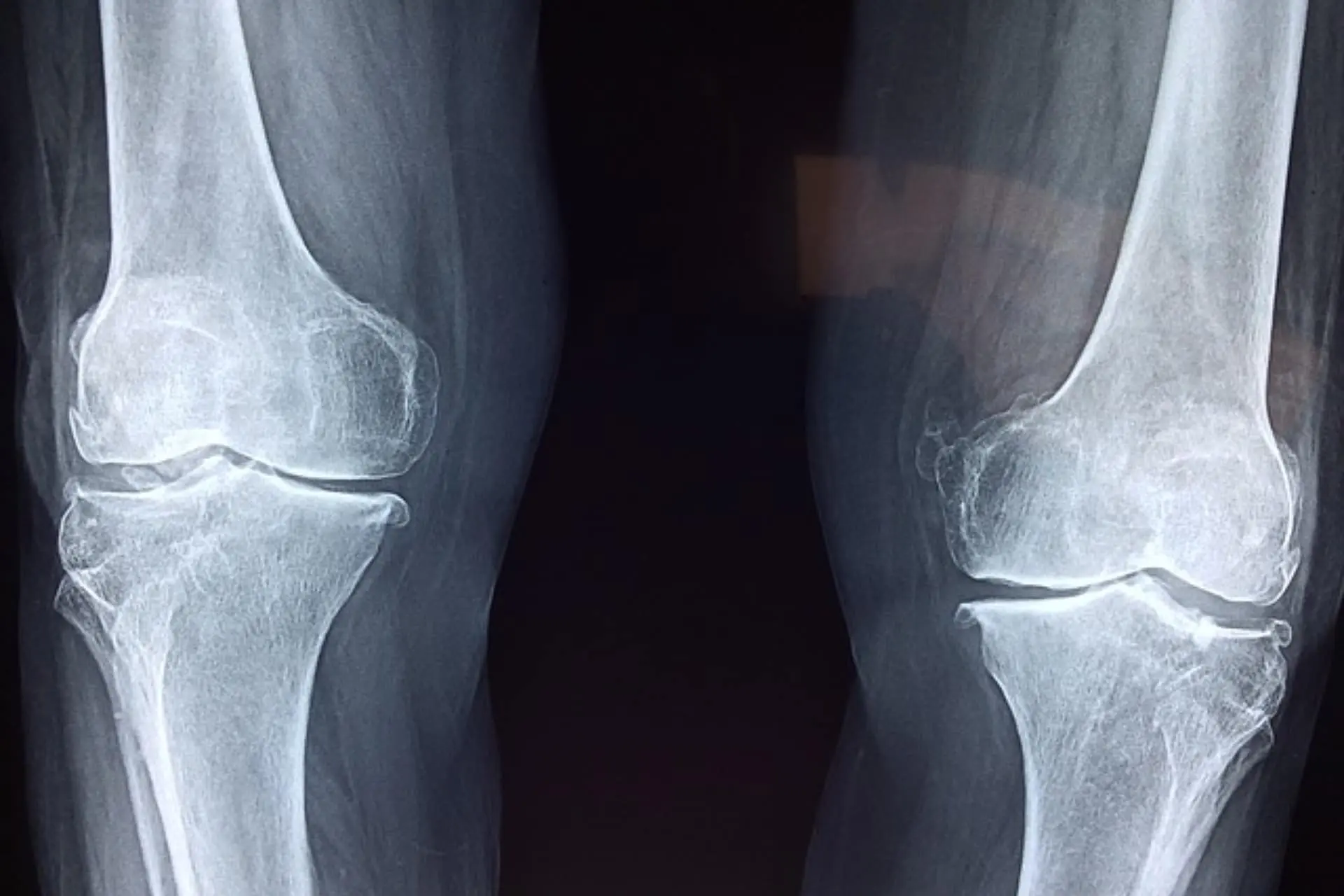
Osteoarthritis is a degenerative disease related to wear and tear of knee cartilage leading to deformity of the bone
LEARN MORE
CAUSES
The most common causes are: trauma, incorrectly consolidated fractures, strenuous work or sporting activities, being overweight, limb deformities (congenital or resulting from trauma).
SYMPTOMS
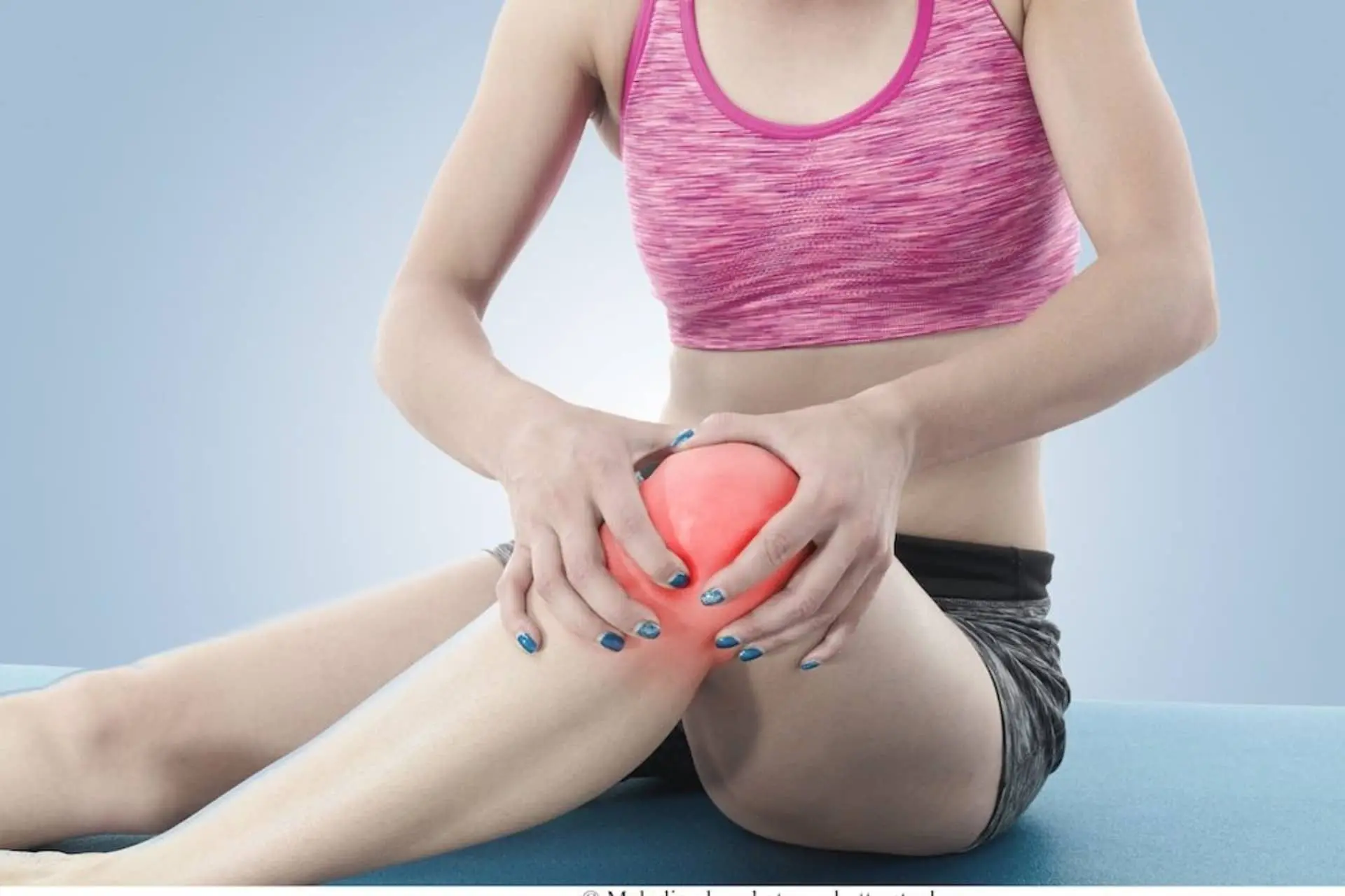
Typical symptoms include pain, stiffness, and swelling of the knee. These symptoms limit the ability to perform even daily activities such as walking, climbing and descending stairs, and usual activities, such as putting on socks and shoes.
PREVENTION
- Maintain an optimal body weight and healthy lifestyle habits.
- Avoid heavy work and excessive loads.
- Nutrition rich in calcium, minerals, and vitamin D.
- Daily physical activity (cycling, swimming).
- Avoid hard surfaces and shoes with soles that are too stiff.
- Avoid prolonged bent position of the knees.
- For preventive purposes, perform one cycle per year of hyaluronic acid and/or PRP infiltrations.
TREATMENT
- Therapy cycles with new generation non-steroidal anti-inflammatory drugs.
- Cortisone treatment for short periods or intra-articular injection in case of strong inflammation or effusion.
- Use specific braces for demanding activities (unloader brace).
- Infiltrative therapy with PRP and/or hyaluronic acid.
- Treatment with stem cells from micro-fragmented adipose tissue.
- In case of failure of these therapies or in advanced cases, surgical management may be needed:
- Arthroscopy and debridement;
- Cartilage transplant ;
- Corrective osteotomy ;
- Joint replacement.
Arthrosynovitis
Athrosynovitis is an inflammatory condition involving the tissue lining (synovium) of the knee
LEARN MORE
CAUSES
The cause is often not known. Some inflammatory diseases of the knee synovium involve only the knee joint. Other diseases, such as rheumatoid arthritis, may affect multiple joints.
TREATMENT
- Anti-inflammatory therapy for 6 months
- “Safe” exercises to improve strength without worsening swelling (e.g., lifting the outstretched leg with a weight on the ankle)
- Ice and application of topical products can be used
- Swelling can sometimes be reduced with the application of elastic stockings and/or braces
- If nonsurgical treatment fails, arthroscopic surgery through which a “synovectomy” can be performed to remove diseased tissue may be useful.
PRECAUTIONS
- Avoid exertion: excessive swelling can weaken the thigh muscle
- Do not ignore or underestimate this condition
- When surgical synovectomy is necessary, keep the limb elevated for 48 hours and start walking under full load 3 days after surgery.
RECOVERY
Because the cause is, in many cases, unknown, recovery may be uncertain.
A good percentage of cases generally recover fully.
Permanent degenerative changes may remain in some cases. Recovery to do common activities after arthroscopic synovectomy is about 3 months.
Knee Arthritis
Knee arthritis is the degeneration of articular cartilage, which causes pain.
LEARN MORE
THREE MAIN TYPES OF ARTHRITIS
- Osteoarthritis (OA): This is the most common of the three. It is a slowly progressive, degenerative disease in which the joint cartilage gradually wears down.
- Rheumatoid Arthritis (RA): This is an inflammatory disease that can destroy joint cartilage, can strike at any age, and generally affects both knees.
- Post-traumatic arthritis: develops after a knee injury, is similar to osteoarthritis, and can develop years after a fracture, ligament, or meniscal injury .
SYMPTOMS
Pain associated with arthritis usually progresses gradually, but sudden onset is also possible.
The joint may become stiff and swollen, making it difficult for the knee to flex and extend. Pain and swelling are greatest in the morning or after a period of inactivity.
The pain may also increase after performing certain activities such as walking, climbing stairs, squatting. The pain can often cause a feeling of weakness in the knee, resulting in a “blockage” of the joint.
Many people experience a change in the level of arthritic pain even as the weather changes.
DIAGNOSIS
Your doctor will perform a physical exam focused on walking, limb range of motion, joint swelling, and tenderness.
Normal radiographs show a decrease in joint space in the affected knee.
Blood tests and other more specific radiological tests, such as MRI, may be necessary for the diagnosis of rheumatoid arthritis .
TREATMENT
In the early stages, knee arthritis is treated with nonsurgical conservative measures.
- Lifestyle modifications that include: weight loss, switching from running to swimming or cycling, and minimizing activities that aggravate the situation, such as climbing stairs.
- Physiotherapy with exercises that increase the range of motion of the joint and strengthen the muscles of the leg and thigh.
- Using supportive tools such as walking sticks, wearing shock absorbing shoes, and using braces can help.
- Other measures may include applications of heat or ice, water exercises, liniments, or elastic bandages.
Several types of medications can be used in the treatment of knee arthritis. Since all patients are different, and since not all people respond to the same medications, your doctor will develop a program for your specific condition.
- Anti-inflammatory drugs, such as aspirin, ibuprofen etc., can be helpful in reducing swelling.
- Glucosamine and chondroitin sulfate as oral supplements can help alleviate the pain of osteoarthritis.
- Corticosteroids, powerful anti-inflammatory agents that can be injected directly into the joint.
- Hyalaronic acid therapy: consists of a series of infiltrations with the aim of changing the characteristics of the joint fluid.
- Special medical therapies for rheumatoid arthritis include the use of gold salts, DMARDs, and other specific medications.
SURGICAL TREATMENT
If arthritis does not respond to conservative treatments.
- Arthroscopic surgery: uses optical fibers to see inside the joint and remove fragments and/or repair cartilage damage
- Osteotomy of the tibia or femur to improve alignment of the knee joint.
- Total or partial knee replacement.
- Cartilage transplant. It is only possible for some knees where cartilage damage is limited.
Orthopedic surgeons are continually searching for new avenues for treating knee arthritis. Research today is focused on new drugs, cartilage transplantation, and other ways to slow the progression of arthritis.
Third Degree Chondropathy
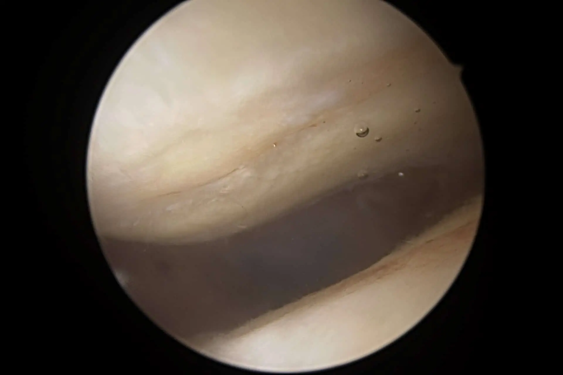
This is a major damage of the articular cartilage of the knee in the loading zone.
LEARN MORE
INJURY
This condition consists of articular cartilage stress that can result in deep fissures that can reach the subchondral layer, and eventually expose the bone itself (grade 4). This condition represents the progression to osteoarthritis in many patients.
CAUSES
The most common cause is abnormally accelerated wear and tear of the cartilage layer of your knee; it may be the result of previous violent trauma (often unrecognized), obesity, or prolonged exertion under load on hard surfaces.
It can also develop as a result of previous trauma or due to loss of the fibrocartilage (meniscus) serving as the shock absorber of the lateral compartment of your knee.
SYMPTOMS
Typical symptoms include pain, which is related to activity under load and is relieved by rest. The pain is often localized to the part where the injury is present.
Slight swelling and weakness are commonly present.
Some patients also experience a sensation of pseudoblocking of the knee.
TREATMENT
- Weight loss, correction of loading or postural abnormalities.
- Regular exercise, but not subjecting the knee to excessive loads (e.g., biking, swimming).
- Anti-inflammatory and/or pain-relieving therapy + viscosupplementation.
- Infiltrative treatment with PRP or mesenchymal cells from adipose tissue.
- Taking dietary supplements such as chondroitin sulfate and glucosamine.
- Walking on soft surfaces with appropriate footwear.
- Arthroscopic surgery to remove damaged and inflamed tissue. Surgical stimulation of degenerated cartilage is successful in cases where the surrounding cartilage is in good condition.
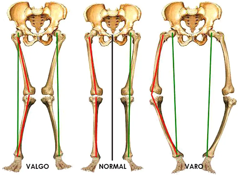
- Surgical correction of the loading axis of the lower limb by osteotomy in varus/valgus knee.
PRECAUTIONS
- No treatment method has optimal success when taken individually.
- You will need to follow all recommendations in order to relieve pain and slow down the progression of cartilage damage.
- Anti-inflammatories should be taken with food. If you continue the therapy for 6 months or more, you should have blood tests to check for side effects.
- Avoid extreme activities involving high pressure on the knee, such as soccer, running, basketball, and tennis.
RECOVERY
Full recovery is rare. Treatment is aimed at improving knee function and delaying the development of osteoarthritis.
In cases treated surgically by arthroscopy, recovery is expected in 3 to 4 months, depending on the extent of damage; in cases of osteotomy, full recovery occurs within 6 months.
Chondromalacia Rotulea
This is often the result of poor conditioning. The patella becomes painful and its underlying cartilaginous surface may be softened.
LEARN MORE
CAUSES
The pain may be dull and continuous, similar to some types of headaches.
The most likely cause of pain is vascular congestion within the patella.
Hormonal factors may also play a role in this condition-in fact, it often occurs in developing girls and then resolves spontaneously after maturation.
Pain may arise as a result of activities involving unusual bending (flexion) of the knee, in walking downhill, or appear after going to the movies and keeping the limb flexed for a long time (so-called theater sign).
SYMPTOMS
The typical symptom is pain, usually in the front of the knee. Pain may be present in both knees. Swelling and stiffness are usually absent.
TREATMENT
- Quadriceps strengthening activities, (e.g., cycling), which must be done regularly for 2-4 months before improvement can be recognized.
- Anti-inflammatory therapy for 2-4 weeks or viscosupplementation and dietary supplementation increase with chondroitin sulfate and glucosamine.
- Weight loss (in case of obesity).
- Local applications of ice, and the use of specific knee pads can also help.
- Walking and bicycling, especially on level ground, are encouraged.
PRECAUTIONS
- Surgical treatment is not indicated as initial treatment for this condition.
- Quadriceps strengthening can increase pain in the first two months-“don’t abandon your exercise program.”
Don’t panic! This condition is common. Recovery is almost always gradual over time
RECOVERY
Estimated recovery time is 6 months, depending on your degree of compliance with the treatment program
Fourth Degree Femoro-Tibial Chondropathy
Breakage, fragmentation, and erosion of cartilage, occurring over time, that has resulted in a full-thickness loss of articular cartilage.
LEARN MORE
CAUSES
The most common cause is abnormal wear and tear of the articular cartilage layer of the knee.
This condition may have developed following severe untreated trauma, excessive body weight, or prolonged loading of the knee on hard surfaces.
It often occurs after a trauma or after the loss of fibrocartilage (meniscus), which absorbs trauma between the tibia and femur.
SYMPTOMS
The typical symptom is pain that increases in relation to activity under load and is relieved by rest. The pain is definitely localized in the area where the lesion is located.
Slight swelling and weakness are commonly present, as well as a slight deficit in knee extension and flexion, and also limping.
TREATMENT
- Weight loss to reduce the load on your knee during walking.
- Exercise regularly such as cycling, and swimming, where the loads on your knees are reduced.
- Anti-inflammatory therapy, physiotherapy, viscosupplementation through infiltrations with the follwing: Hyaluronic acid, glucosamine, PRP, mesenchymals from adipose tissue.
- Walk on soft surfaces and wear comfortable shoes; the use of a brace that corrects the load axis can decrease pain.
- Arthroscopic surgery is useful for removing damaged and inflamed tissue and for surgically stabilizing the cartilage defect. Surgical stimulation of the degenerated cartilage can lead to positive results.
- An open surgical procedure, which includes cutting the bone (osteotomy) may be necessary to straighten the lower limb in the most severe deformities.
PRECAUTION
- Avoid physical activities involving excessive knee stress, such as tennis and running.
- Anti-inflammatory drugs should always be taken after lunch or dinner. If you prolong the anti-inflammatory therapy for 6 months, you will have to undergo blood tests to check that there are no side effects.
- Your activities and your diet must be aimed at optimizing bone density; moderate exercise is usually more useful if done daily.
- No single treatment has optimal efficacy. You will need to follow all recommendations in order to relieve pain.
RECOVERY
A full recovery is rare; the treatment was designed to improve knee function and delay the need for a knee replacement as much as possible.
In cases treated only with arthroscopic surgery, recovery is gradual, over a period of 3-4 months.
If an osteotomy has been performed (cutting the bone to correct the alignment), crutches are generally used for at least 1 month, and full recovery occurs within 6 months.
Jumper's Knee
LEARN MORE
CAUSES
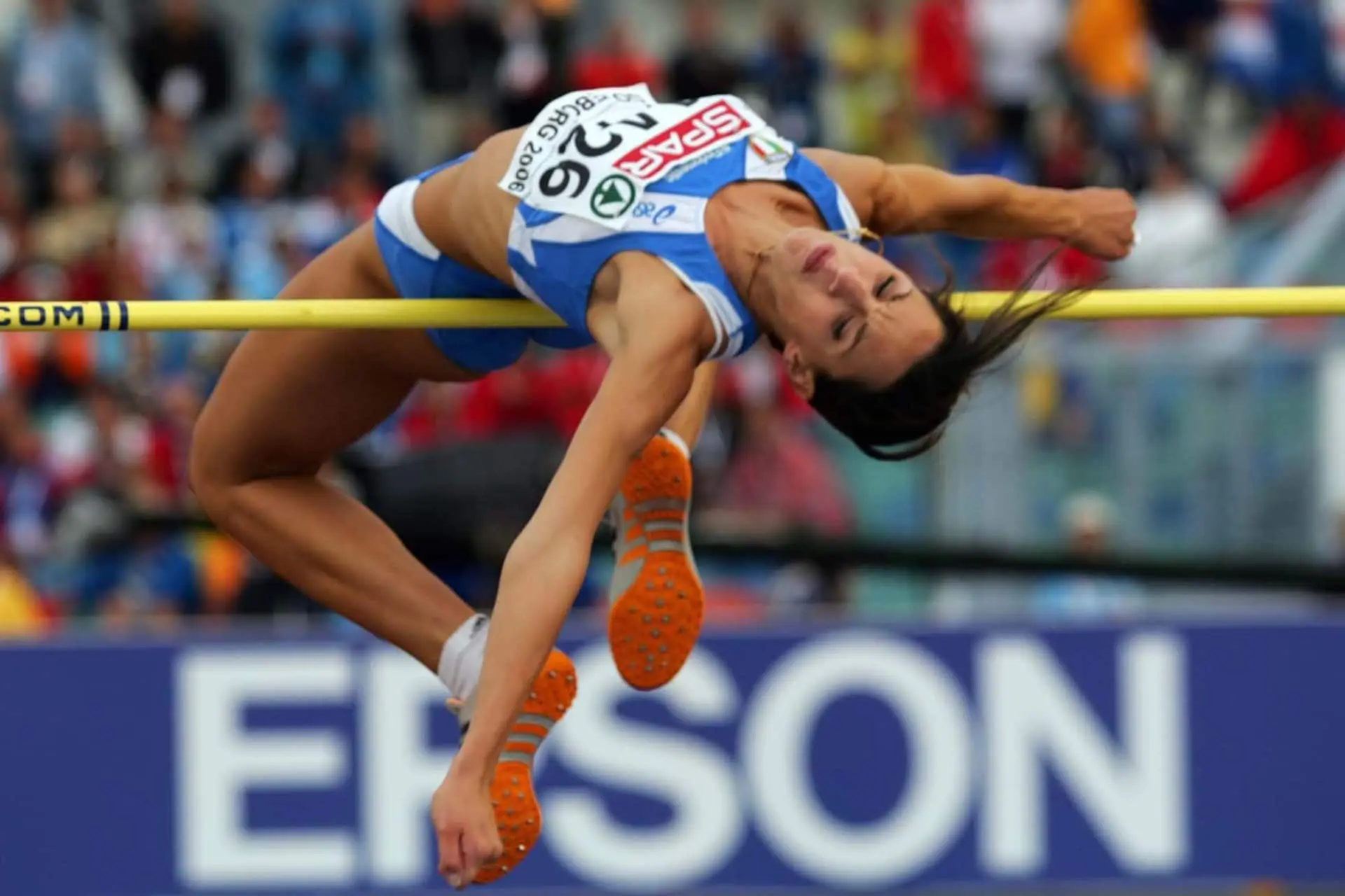
The most common cause is repeated micro-trauma to the tendon during activities involving prolonged running or jumping.
SYMPTOMS
Typical symptoms are: pain at the lower pole of the patella, aggravated by activities such as jumping or running. In severe cases, swelling also appears. Associated quadriceps weakness is an unfavorable sign.
TREATMENT
- Anti-inflammatory therapy for 1-2 months.
- Use of ice at least 3 times a day.
- A knee brace or compression bandage can relieve the pain.
- Physical activity should be modified in order to reduce stress on the patellar tendon.
- A technique involving multiple punctures with a needle tip may be employed by your physician to stimulate spontaneous healing
- In cases that have proven resistant to treatment for 1 year, minor surgery is required to remove necrotic tissue
PRECAUTION
- Avoid cortisone injections as it weakens tissue and can lead to more damage and tendon rupture.
- Be patient, unfortunately, improvements in this condition are almost always very gradual.
- Control your diet; high-fat diets can be deficient in important minerals and vitamins.
- Smoking can also contribute to this condition.
- Avoid squatting on hard surfaces.
RECOVERY
the expected recovery time is slow. More tenacious cases may require minor surgery. Improvement is gradual, taking a minimum of 6 months.
Knee Gout
Gout is a dysmetabolic disease that results in increased uric acid in the blood, with subsequent deposition of uric acid crystals in peri-articular tissues
LEARN MORE
INJURY
Accumulation of the deposits in turn causes very painful inflammation in the joint and can evolve into chronic deforming forms of arthritis.
In addition, deposits can also form in the kidneys, causing kidney stones, or in the subcutaneous tissue with the formation of nodules (tophi).
Kidney colic caused by calcinosis can also present without previous joint symptoms.
SYMPTOMS
The acute gout attack may resolve spontaneously, but generally lasts 7 to 10 days.
It can affect several joints including the knee. The ‘joint becomes painful, red and swollen.
TREATMENT
- Generally, in the acute phase, drug therapy is intervened with avoiding the intake of drugs that could aggravate the condition (diuretics, aspirin, nicotinic acid) and alcohol; the patient should also take bed rest
- As for chronic pathology, action is generally taken in cases of hyperuricemia (level above 13 mg/100cc in men and 10 mg/100 cc in women), uricosuria (uric acid present in urine in amounts above 1,100 mg/day), frequent acute attacks, nodules, gouty arthropathy, renal stones, or renal damage. In these cases, therapy will almost always have to be followed for life and will be based on the individual patient’s status.
- It is a good idea to follow a diet with a normal protein content, not exceeding in sugars and limiting alcohol consumption as much as possible.
- In particular, alcohol and obesity are factors that increase uric acid secretion and decrease its elimination.
RECOVERY
In acute attack, the onset is dramatic, with pain in the very early morning hours, that is, when blood stasis is greatest. The mere weight of blankets can be unbearable.
We have hyperemia (redness), tumefaction (swelling) and local rise in temperature and also fever. Usually the crisis stops after a few weeks but may recur and become increasingly frequent.
In order to prevent the onset of gout, it may be necessary to follow an appropriate diet:
- Avoid foods high in purine bases (sweetbreads and offal, fatty meats and fish, cured meats, shellfish);
- Limit protein intake to one gram per day, per kilogram of ideal body weight;
- Drastic reduction in dressing fats;
- Drink plenty in order to ensure good diuresis;
- Limit diuretics, which, by increasing urate excretion, can promote nephrolithiasis.
Multidirectional Knee Instability
The result of complete rupture of at least two of the most important ligaments in your knee including anterior and posterior cruciate ligament. This combined rupture causes significant instability in the knee, which can be felt even during normal daily activities. There may be associated damage to the cartilages of the knee.
LEARN MORE
CAUSES
The most common cause is knee trauma that can be torsional, hyperextension, or lateral flexion. The knee may be exposed to violent forces during ski jumping, collisions during sports, or major traffic accidents.
SYMPTOMS
Typical symptoms are immediate disability, followed by severe pain and swelling within the first 12 hours.
TREATMENT
- Ice and elevation of the limb to limit swelling.
- Observation of pulses to ensure that circulation is intact.
- Anti-inflammatory and pain-relieving therapy is helpful.
- Use of crutches and a knee brace.
- Generally, the solution is surgical, after the acute symptoms have disappeared (usually 1-6 weeks after the injury).
- Physical therapy is helpful to reduce swelling and stiffness, improve muscle control, and decrease pain.
PRECAUTIONS
- Limit swelling, both at the knee and distally at the foot and ankle. Use ice and keep the knee elevated above the level of the heart until swelling improves.
- Check for signs of inadequate circulation in the foot, such as pallor or reduced pulse. Report any signs to your doctor immediately.
- Although surgery is generally necessary, it should not be undertaken while the knee is still in a state of significant stiffness and inflammation.
- Do not neglect your home program of therapeutic measures (ice, rest, limb unloading, and medical therapy).
RECOVERY
Expected recovery after surgical repair of multiple ligament rupture can be long, about 6-12 months.
Guided physical therapy is often prescribed for a period of 2-3 months, followed by an independent exercise program.
Unfortunately, you have suffered one of the most serious knee injuries.
Medial Collateral Injury
Three grades of injury are distinguished in order of severity. Grade 1 and 2 are treated bloodlessly. Only in the most severe case is surgical indication placed.
LEARN MORE
CAUSES
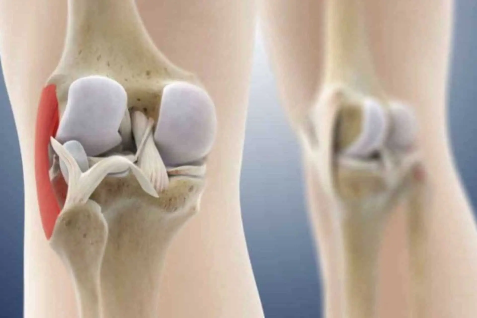
Isolated medial collateral ligament (MCL) injury typically occurs from a combined flexion, rotation, and valgus motion of the knee.
The LCM has a superficial and a deep layer that has connection with the medial meniscus and conditions its movements.
SYMPTOMS
Isolated medial collateral ligament (MCL) injury typically occurs from a combined flexion, rotation, and valgus motion of the knee.
The LCM has a superficial and a deep layer that has connection with the medial meniscus and conditions its movements.
TREATMENT
- Ice, limb unloading, and elevation with semi-flexed knee relieve pain.
- Anti-inflammatory therapy immediately after the injury is useful.
- Application of functional bandage with LCM support and possible brace with 30°-90° joint range limitation.
- Partial load on the limb for 7 days in grade 1 injuries and 15 days in grade 2.
- Surgical treatment in severe cases and when other injuries are associated.
PRECAUTION
- Begin treatment immediately with ice and limb unloading.
- Do not minimize the injury because the MCL has an important stabilizing function in the knee during the performance of many sports and work activities. Inadequately treated injury can create permanent damage to the knee.
- Avoid hyperextension and external rotations for at least one month.
- Adopt preventive brace or bandage for risky sports (skiing, motocross, soccer, basketball, tennis).
RECOVERY
Full recovery after bloodless treatment occurs in about 30 to 40 days. In more severe cases treated surgically, about 3 months are required before returning to sports.
ACL injury
Sprain injuries with ACL injury are a frequent occurrence in many sports (soccer, motocross, basketball, skiing) in women the frequency is about 3 times higher than in men
LEARN MORE
CAUSES
The ligament can tear due to a torsional movement, a hyperextension of the knee, or an abrupt contraction of the quadriceps with the knee flexed as occurs from violent compression with skis.
SYMPTOMS
Typically, the subject experiences a popping noise and the sensation that the knee is coming out of its socket; the pain is not always so violent and the injured person is able to continue but immediately experiences a feeling of instability.
The knee tends to swell prematurely.
TREATMENT
- Ice, discharge and elevation of the limb.
- Anti-inflammatory and pain-relieving therapy.
- Use of crutches and knee brace.
- Generally the solution is surgical: wound repair or reconstruction .
- In some special cases, non-surgical treatment can be performed.
PRECAUTION
- Limit swelling by using ice and keeping the limb elevated.
- Initiate home treatment after the first week to regain knee mobility.
- Make sure by medical examination and possible magnetic resonance that there are no associated meniscal or cartilage lesions.
- Do not neglect the injury you have sustained as it could be the “beginning of the end” of your sporting activity.
RECOVERY
The anterior cruciate ligament reconstruction surgery performed under arthroscopic control involves a long rehabilitation during which your will and commitment is of fundamental importance.
The common activities of relationship life (walking, driving the car, working in the office) are resumed after about 1-2 months.
For low-risk sports (swimming, cycling, golf, jogging) it takes about 3 months while for the more dangerous ones it takes 6 months.
LCP injury
This injury consists of a complete rupture of the ligament that stabilizes the posterior translation of the tibia on the femur
LEARN MORE
INJURY
The posterior cruciate ligament is located centrally within the knee just, behind the anterior cruciate ligament, making it difficult for the patient to locate the affected area.
CAUSES
The most common cause is a severe blow to the front of the tibia, with the knee in a bent position (such as in hitting the dashboard of the car in a car accident).
SYMPTOMS
Typical symptoms are swelling of the knee with signs of instability, but rarely are there true episodes of knee failure. Pain may be minimal.
TREATMENT
- Ice and elevate the limb to reduce swelling.
- Flexion-extension within the limits allowed by pain is encouraged
- Cycling should be started as soon as an adequate degree of moblity is reached.
- Quadriceps strengthening exercises, such as the squat from a standing position (with elevation on the toes) and the leg press, are recommended.
- Exercises for the flexors should begin only later.
- Most cases are treated conservatively with the help of a special brace.
PRECAUTIONS
- Do not participate in aggressive sports until you have achieved good quadriceps tone and the swelling has disappeared.
- Be sure to fully understand your rehabilitation program. This is essential for optimal recovery from this injury.
RECOVERY
Usually, recovery is expected in about 3 to 4 months, at which point, swelling should be gone and strength recovered. A full return to sports activities is usually possible.
Meniscal Injury
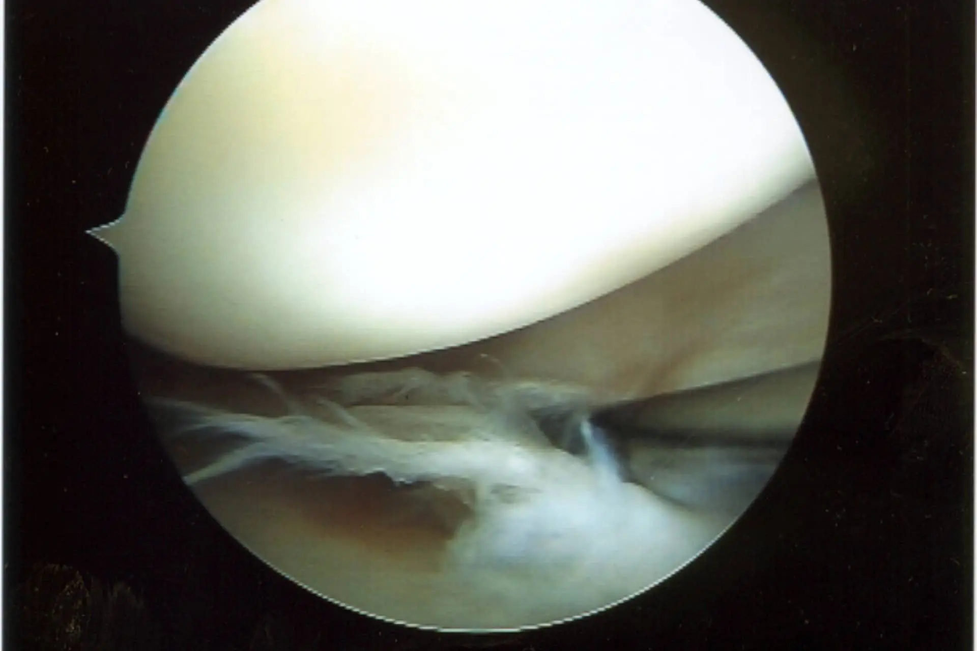
This condition consists of a tear in the fibrocartilage (meniscus) deputed to absorb trauma and lubricate the inner or outer compartment of the knee under load.
LEARN MORE
CAUSES
The most common cause is excessive pressure applied to the knee fibrocartilage. Usually the injury is created, during trauma with twisting or excessive flexion of the knee (sometimes the patient experiences a cracking noise).
SYMPTOMS
Typical symptoms are: localized pain in the area of knee injury, usually sharpened by exertion and the act of crouching. One may also experience mechanical sensations of popping and jamming up to joint locking when the medial meniscus ruptures “bucket-handle.”
TREATMENT
- Observation of symptoms, spontaneous recovery is rare in adults, meniscus should be saved when possible.
- When symptoms are minimal practice safe activities to strengthen muscles (e.g., bicycling) that avoid twisting and excessive bending movements.
- Optional anti-inflammatory therapy for 1-2 weeks.
- Arthroscopic surgery to adjust or repair the damaged meniscus. In some cases, partial regeneration of fibrocartilage tissue may occur after surgical stimulation with radiofrequencies.
PRECAUTION
- In nonsurgical treatments, sports activities involving significant soreness should be avoided.
- Anti-inflammatory therapies are a measure to be taken only temporarily, and not for a prolonged period .
- Cortisone injections should be avoided because they can mask the real condition and thus lead to more damage.
RECOVERY
Recovery after arthroscopic regularization of the damaged meniscus (partial meniscectomy) is estimated in 1-4 weeks, in case of meniscal suture a brace should be adopted for 15 days avoiding full load on the limb and full knee flexion will be prohibited for the first month. Sports recovery usually occurs after 3 months.

Limited Knee Extension
Limited ability to fully straighten the knee, often caused by injury, arthritis, muscle imbalance, or mechanical blockage such as meniscus injury.
LEARN MORE
CAUSES
The most common cause is the presence of scar tissue and swelling around the patella. Secondarily, shortening of the thigh and calf flexor muscles can also limit extension, as can ligamentous reconstruction with suboptimal graft placement.
SYMPTOMS
Typical symptoms include pain in the anterior compartment of the knee; thickening of tissue below the patella and quadriceps atrophy may also be present.
TREATMENT
- Anti-inflammatory therapy and FKT for 1-2 months.
- Patellar mobilization: sit with your leg extended as far as possible and relaxed. Move your kneecap firmly in various directions for 5 minutes.
- Seated extension: sit on table or floor with legs straight. Rest your feet on an elevated support so your knee remains suspended, unsupported, place a weight over your kneecap and contract.
- Extension from prone position: lie on your stomach, with feet and ankles protruding over the edge (of the bed, or a table). Relax and allow gravity to straighten the leg. To increase the stretch, place a light weight around the ankle. Hold the position for 5-10 minutes,
- Long stride walking: increase the distance of your walks and also increase the stride length as much as possible.
- Exercise regularly, change your sleeping position by trying to sleep supine with your legs extended.
PRECAUTION
- Hold each stretch for 15 to 30 seconds. Do not bounce or jerk suddenly during stretching.
- Ice on the knee for 15-20 minutes after stretching.
- Stretch until you feel moderate tension or pain. Frequent repetition of exercises is much more useful than brute force!
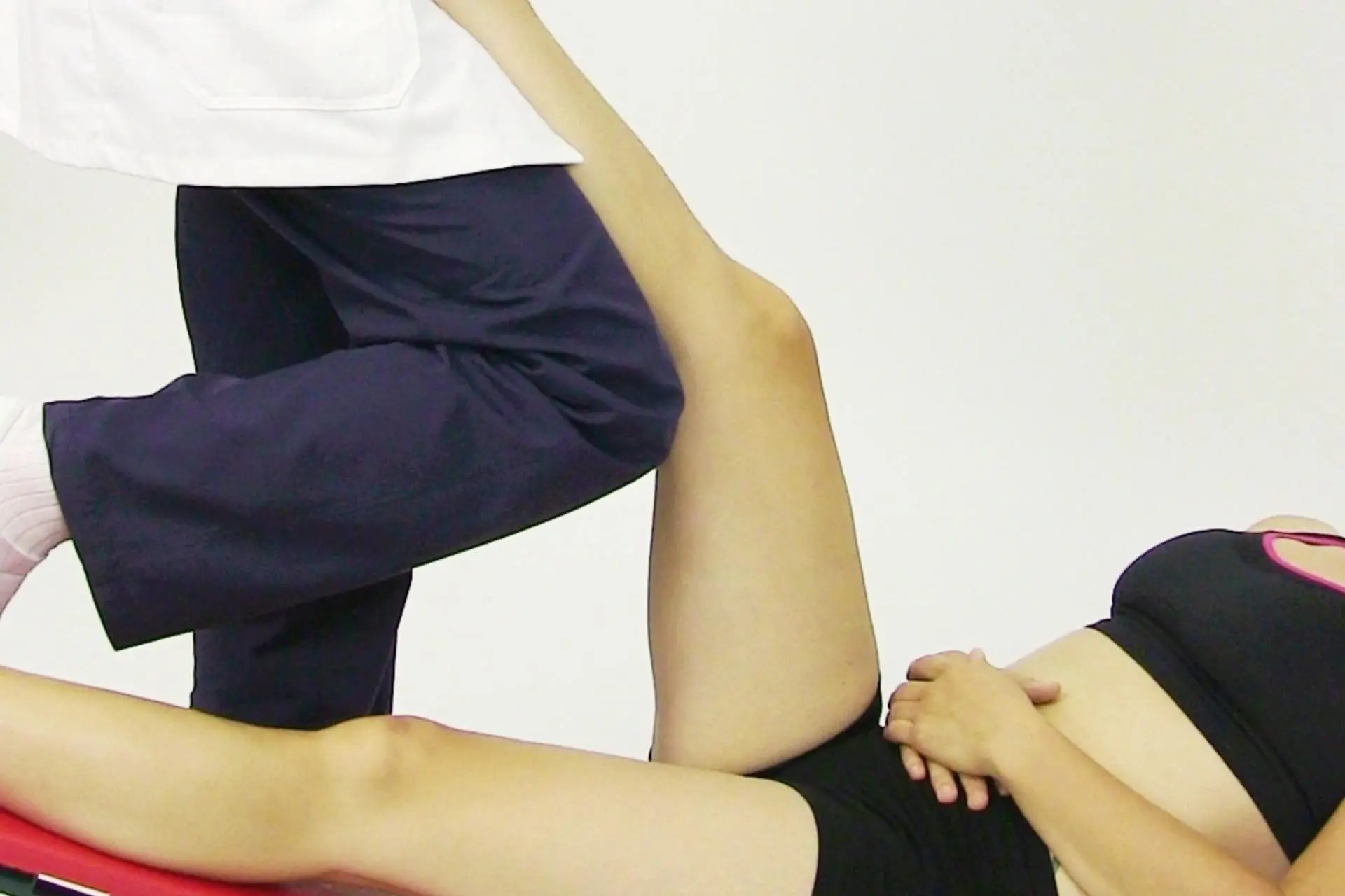
Limited Knee Flexion
Limited knee flexion is the limited ability to fully bend the knee, often caused by injury, swelling, arthritis, or soft tissue tension.
LEARN MORE
CAUSES
The most common cause is excessive tension (contraction) of the connective tissue around the patella that prevents it from sliding. Scarring in the aftermath of previous trauma or surgery can give rise to this condition as well.
SYMPTOMS
Typical symptoms include inability to fully flex the knee and anterior compartment pain. The tissues are atrophied and the flexor muscles are relatively weak.
TREATMENT
- Regular intake of anti-inflammatory drugs and FKT for 1-2 months.
- Patellar mobilization: sit with your leg fully extended and relaxed. Move your kneecap firmly in all directions for 5 minutes. You can also ask a friend to perform these maneuvers.
- Sliding on the wall: lying on your back with your legs flexed at 90° and your feet resting on the wall. By gravity, let your feet slide down the wall as far as you can reach (use a sock to reduce friction). You can help by pushing with the other leg as well.
- Squats from the chair: sit in a chair with your arms at rest, along your sides. Your feet are firmly resting on the floor. Glide forward with your legs and pelvis, balancing your weight with your arms.
- Kneeling Squats: on all fours, with hands and knees on the floor. Stretch your back, bringing your pelvis toward your feet. When you feel pulling, hold the position as long as possible.
- Bicycle: You can use a bicycle to gain flexion. Position the saddle at a height that allows you to pedal without having to get off the saddle. Progressively lower the saddle to achieve greater knee flexion.
- Practice regular physical activity as additional therapy.
PRECAUTION
- Stretches to the point where tension or moderate pain is felt.
- Do frequent repetitions and avoid brute force.
- Hold each stretch for 15 to 30 seconds. Do not bounce or jerk suddenly during stretching.
- Ice on the knee for 15-30 minutes after stretching.
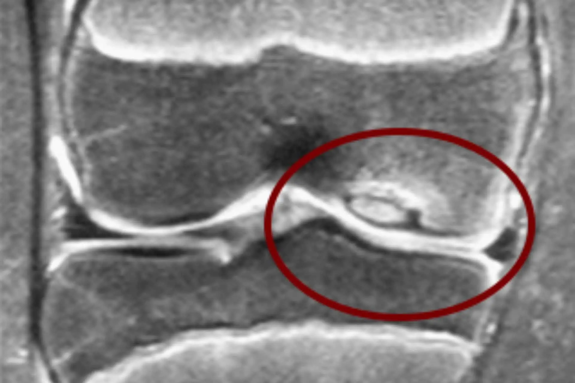
Osteochondritis Dissecans
Osteochondritis of the knee is a disease process characterized by the partial or complete detachment of a chondral or osteochondral fragment from the articular surface of the condyles.
LEARN MORE
INJURY
If the fragment falls into the joint, a free body is formed and we speak of osteochondritis dissecans. In most cases the medial femoral condyle is affected (85 percent), then the lateral femoral condyle (13 percent) and finally the anterior surface of the trochlea (2 percent).
Based on the age of onset, we can roughly distinguish a juvenile osteochondritis dissecans, which strikes in an age group generally between 10 and 16 years old, and an adult osteochondritis dissecans.
SYMPTOMS
The typical symptom is pain with episodes of pseudo-blockage accompanied by tumefaction. The clinical picture is essentially poor; often, chondral or osteochondral lesions detected on MRI or by arthroscopy are asymptomatic and thus represent an occasional finding.
There are no specific clinical trials for this type of pathology.
Clinically, it is possible to detect generic effusion, accompanied by pain on palpation of the affected hemirima, joint rocking, and limitation of articularity.
The only test that can guide is Wilson’s: the knee is sore if you extend it intra-rotating the tibia at the same time.
However, it is the instrumental examinations that allow a diagnosis of certainty
TREATMENT
The prognosis is good, especially in children, where there is complete healing in most cases; in dissecting forms it is sometimes possible to reinsert and fix the fragment.
In adults, the issue is maintaining an intact joint surface; in fact, cartilage or osteo-cartilage damage is considered to be pre-arthritic degeneration.
Treatment, whether conservative or surgical, depending on the subject’s age, site, and extent of injury aims to restore normal articular cartilage in order to delay the onset of the arthrotic degenerative process as much as possible.
RECOVERY
Recovery time is related to the age of the patient and the type of injury. Children generally recover quickly even with bloodless treatment. In surgically treated adults, recovery is gradual and may take more than 6 months.
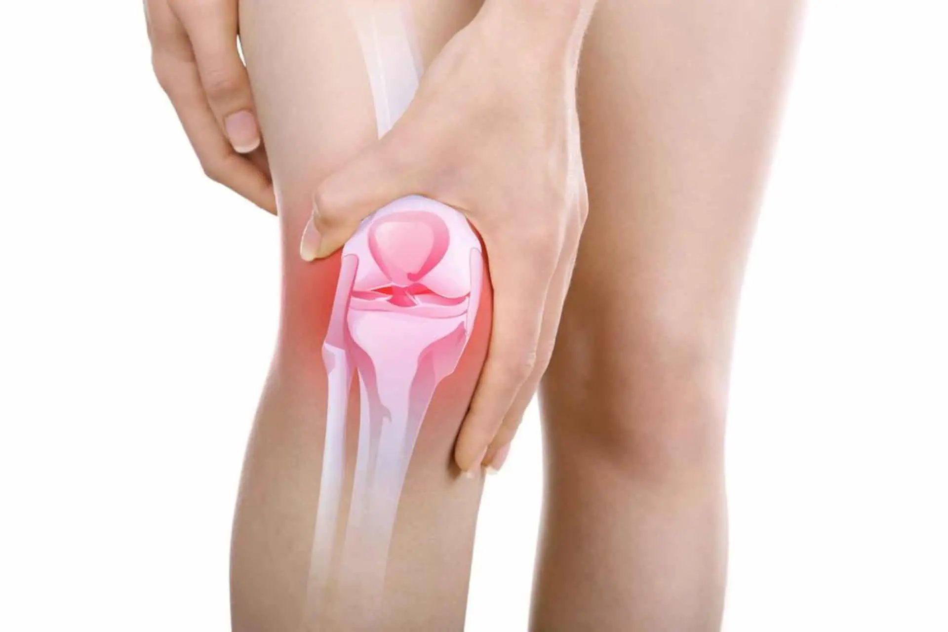
Patellofemoral Joint
Patellofemoral pathology includes a number of conditions that affect the patellofemoral joint, causing pain in the front of the knee.
LEARN MORE
PATELLAR FEMORUS
The patellofemoral joint is extremely important for being able to perform many normal activities, such as walking, running, climbing and descending stairs. It consists of the femoral trochlea and patella, which allows flexion-extension of the knee and any flexion-extension movement of the leg against resistance. The patella has the function of centering during the contraction of the quadriceps with the effect of increasing the force and braking the knee flexion movement, as well as distributing the forces applied to the patellar tendon and stabilizing the knee in rotational movements. Patellofemoral pathology represents a cause of suffering and disability. It is initially manifested by joint discomfort, difficulty kneeling and squatting, and pain especially when walking downhill.
CAUSES
There can be many causes of patellofemoral pain, which also occur at a young age, especially in women. Most patellar syndromes are the consequence of impaired functioning of the knee extensor system, which includes various muscular, tendon, ligament and osteo-articular structures.
Any imbalance of the forces acting on the patella can lead to the onset of painful syndromes.
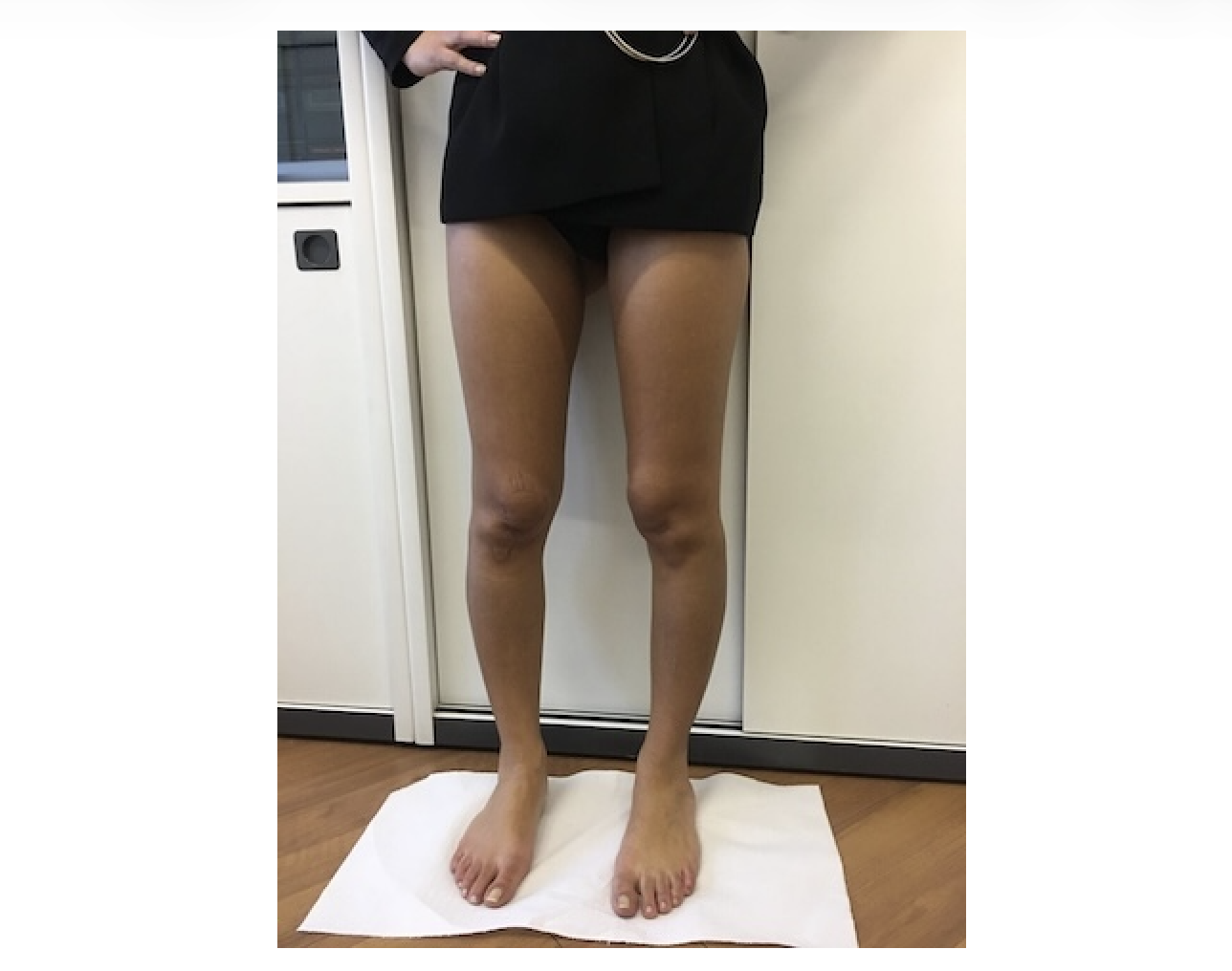
Determining factors may be the reduction of quadriceps tonotrophism, the presence of valgus of the knees with an increased femoro-tibial angle (called “Q” angle), excess intra-femoral rotation, patellofemoral dysplasias, ligamentous hyperlaxia, and any other situation involving altered joint biomechanics.
The quadriceps muscle plays a fundamental role in patellar stability and in particular, the vastus medialis oblique ensures vertical, horizontal and rotational stability.
Frequently the cause is an overload of the extensor apparatus in the presence of determining factors, for example work activities on the knees. Sometimes an acute traumatic episode can trigger symptoms.
Most patellofemoral syndromes can greatly benefit from conservative treatment and good results are reported in more than 60% of cases.
TREATMENT
When the ailment is mild and transient, the best cure is to rest and avoid activities that cause pain, this can mean not squatting or kneeling, avoiding prolonged positions with bent knees (like in the cinema) or avoiding long walks downhill.
Nutritional therapy, the adoption of orthotic insoles and the constant practice of specific motor activities may help to achieve a remission of symptoms.
In our experience, a good rehabilitation program including postural gymnastics, stretching and selective muscle strengthening, associated with hyaluronic acid therapy, can definitively resolve most of these cases.
If the patella is subluxing, especially in young people, it may be advisable to use a neoprene knee brace that stabilizes the patella during the most demanding activities.
An important role in the treatment of patellofemoral syndromes is represented by bio-orthopaedics, and in particular the use of PRP.
If conservative treatment fails, surgical treatment may be necessary, aimed at restoring normal joint mechanics (realignment) and treating any cartilage damage.
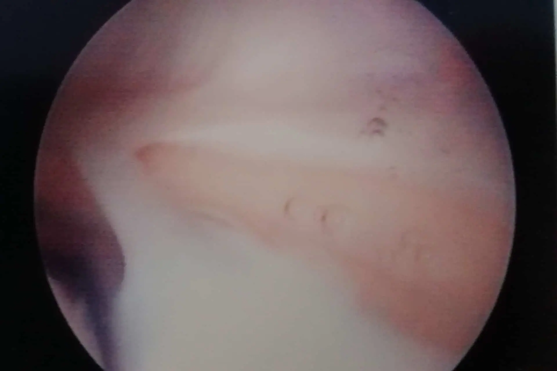
Synovial Plica And Pseudo Blockade
Synovial plica is one of the most common causes of transient knee pain; it consists of the presence of an abnormally thickened membrane (“plica”) within the knee that can result in pain and difficulty in movement.
LEARN MORE
CAUSES
These disorders are usually caused by previous trauma, often missed, or joint wear and tear. Because the knee is loaded, with full body weight, 1 to 4 million times a year in active people, spontaneous recovery may not always occur.
SYMPTOMS
Typical symptoms are recurrent, low-intensity pain in common activities and sometimes there may be knee crepitus and/or pseudoblocking.
TREATMENT
- Activity modification; e.g., replace jogging with bicycling.
- Observation only for 3-6 months to allow for slow natural recovery.
- Anti-inflammatory therapy for several weeks or viscosupplementation for 1 month.
- If there is a particularly sensitive knee area, apply ice after physical activity.
- In persistent cases, arthroscopic surgery may be necessary to remove the abnormal tissue and clean the joint.
PRECAUTIONS
- Stretches to the point where tension or moderate pain is felt.
- Do frequent repetitions and avoid forced movements.
- Hold each stretch for 15 to 30 seconds. Do not bounce or jerk suddenly during stretching.
- Ice on the knee for 15-30 minutes after stretching.
Ileo Bendellectomy Friction Syndrome
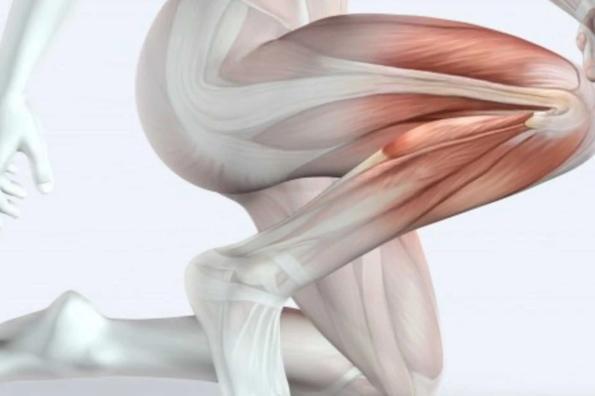
This condition consists of persistent inflammation with pain at the level of the external condyle of the femur exacerbated by flexion-extension movements of the leg
LEARN MORE
CAUSES
The most common cause is “rubbing” of the iliotibial band, on the femoral condyle due to “overuse” of the ‘limb as occurs, in endurance running or cycling when undergoing excessive training. Any attempt to continue, despite the pain, results in increased inflammation.
SYMPTOMS
Typical symptoms include pain, which usually appears during a downhill run or walk and radiates from the lateral compartment of the knee. There is almost never visible swelling.
TREATMENT
- Rest from jogging, running or mountain walking until you can perform these activities without pain.
- Anti-inflammatory therapy for 1-3 weeks.
- Local application with ice. Band stretching exercises.
- Limit the local use of cortisone, which can mask pain, and thus lead to a greater risk of “abuse”.
- The surgical solution is used only in case of failure of bloodless treatment.
- PRP injection
PRECAUTION
- Don’t try to keep running despite the pain.
- Avoid surfaces slightly inclined towards the affected side of your knee.
- Unnecessary orthopedic corrections (e.g. orthopedic insoles) can contribute to the onset of this condition
- Change your sporting activities.
- Make sure your running shoes absorb shock well during heel strike.
RECOVERY
The time needed to achieve complete recovery can be several weeks.
Patellar Hyperpressure Syndrome
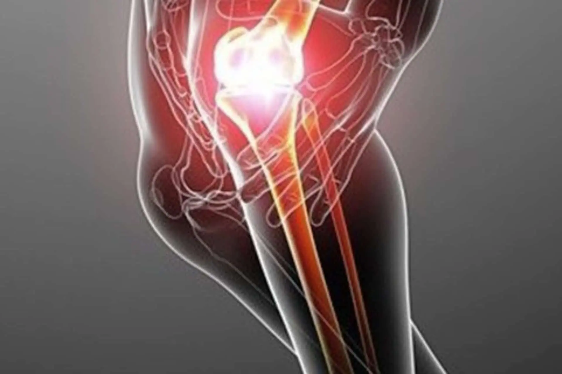
This condition is caused by excessive patellar pressure on the external facet that, over the years, leads to premature erosion of the patellar cartilage.
LEARN MORE
CAUSES
The most common cause is abnormal tension of the external wing ligament, which results in hyperpressure on the lateral part of the patella.
SYMPTOMS
Typical symptoms are a creaking sensation and pain during activities involving knee flexion, such as descending stairs. Mild swelling and increased tenderness along the outer edge of the patella are also common.
Prolonged flexed limb posture such as sitting in a movie theater can increase discomfort.
TREATMENT
- Anti-inflammatory therapy for 2-4 weeks.
- Avoid activities that require deep knee bends.
- Long walks on level ground and exercises for the quadriceps femoris, such as extended leg lifts with light ankle weights, can be helpful.
- Weight loss reduces pressure on cartilage.
- An elastic knee pad stabilizing the kneecap can provide relief.
- Shark cartilage, chondroitin sulfate, glucosamine viscosupplementation with Hyaluronic acid can help cartilage healing.
- Most often there is benefit through arthroscopic surgery, in which the overstretched ligament is loosened (lateral release) and the damaged cartilage is regularized.
PRECAUTION
- Episodes of repeated swelling, even if mild, often indicate worsening cartilage damage.
- Long-term anti-inflammatory therapy is not indicated for this particular condition.
- Sporadic physical abuse, such as downhill walking, can accelerate permanent cartilage damage.
- Arthroscopic surgery is necessary; it is best done before major cartilage damage is created.
RECOVERY
With medical therapies, gymnastics and observing the above, gradual improvement can be observed over 3 months.
If arthroscopy has been performed, ambulation with a brace/bandage is prescribed for the first week, followed by a rehabilitation program that includes swimming and cycling.
Return to competitive sports can take up to 4 months.
Femoro-Rotulea Syndrome
Patellofemoral syndrome is a condition that causes pain in the front of the knee due to improper movement of the patella.
LEARN MORE
SYMPTOMS
The pain, which can be very significant, arises suddenly, sometimes for no reason, also leading to an antalgic joint blockage.
DIAGNOSIS
Conventional radiology, performed with the knee in flexion, aids in diagnosing the syndrome in most cases. Magnetic resonance imaging (MRI) can be done in cases where diagnosis is more difficult, or when a possible cartilage injury is suspected. CT is also used in the study of this joint.
CAUSES
To understand the nature of the problem, we must understand the mechanical reason for it.
The pain is related to inflammation of the cartilage of both the patella and the femoral condyles (chondropathy).
This inflammation is linked to poor patellar sliding during knee flexion-extension. In particular, the patella becomes “lateralized,” meaning it slides more on the outer edge of the knee, increasing friction with the femur.
Such “lateralization” also called “external overpressure” or “patellar malalignment” can be mainly due to 3 situations:
Quadriceps Atrophy
Functional Overload in Knee Flexion
Anomaly of Femoral Gutter Anatomy
Quadriceps atrophy is the major cause leading to this syndrome (90% of patients); the stabilizing strength of the muscle opposing lateralization of the patella, is lacking.
Other times, there may be a cause related to overexertion, as in those patients in whom pain is triggered after a “walk” in the mountains, or a heavy sports session, especially if one has played a sport with the knee flexed (e.g., volleyball).
In a very small proportion of patients, the problem may instead be due to an anatomical abnormality of the femoral gutter, which is “flared” and thus poorly contains the patella.
These patients also typically have a history of repeated patella “dislocations” with recourse to painful “reductions” in the emergency room.
TREATMENT
Treatment of patellofemoral syndrome is, in most cases, physiotherapy. Careful functional reeducation of the quadriceps is necessary, especially developing the vastus medialis oblique, which is an important muscle.
The patient will conclude the re-education with freestyle swimming (not breaststroke) and high-saddle bike (soft). The use of special patellar knee braces may be recommended on the recommendation of the orthopedic specialist.
Rarely, and with sometimes unsatisfactory results, treatment is surgical. Surgery is in fact used rarely in patients who have dislocated the patella in at least one episode.
The current surgical treatment is arthroscopic with a combined Lateral Release and Medial Capsular Retention procedure, etc. Halbrecht.
In this type of surgery, sectioning of the external wing ligament that “pulls the patella outward” and retention with an all intra-articular suture of the medial capsule that is lax is done.
Other surgeries such as transposition of the patellar tendon insertion are now used increasingly rarely and in very selected cases, given the invasiveness of the procedure.
Among these, the one that has shown the best results is the one described by Fulkerson.
Goosefoot Bursitis
Goosefoot bursitis is an inflammation of the goosefoot bursa, which causes pain on the inside of the knee.
LEARN MORE
SYMPTOMS
The so-called goose leg is located at the level of the proximal portion of the tibia on the inner side of the leg where the tendons of the semitendinosus, gracilis and sartorius are inserted. This area is covered by bursal tissue that facilitates sliding but in case of inflammation it can cause bursitis and pain in the movements of external erotation of the leg, flexion and valgization.
Pain can also be present at rest and disturb sleep.
DIAGNOSIS
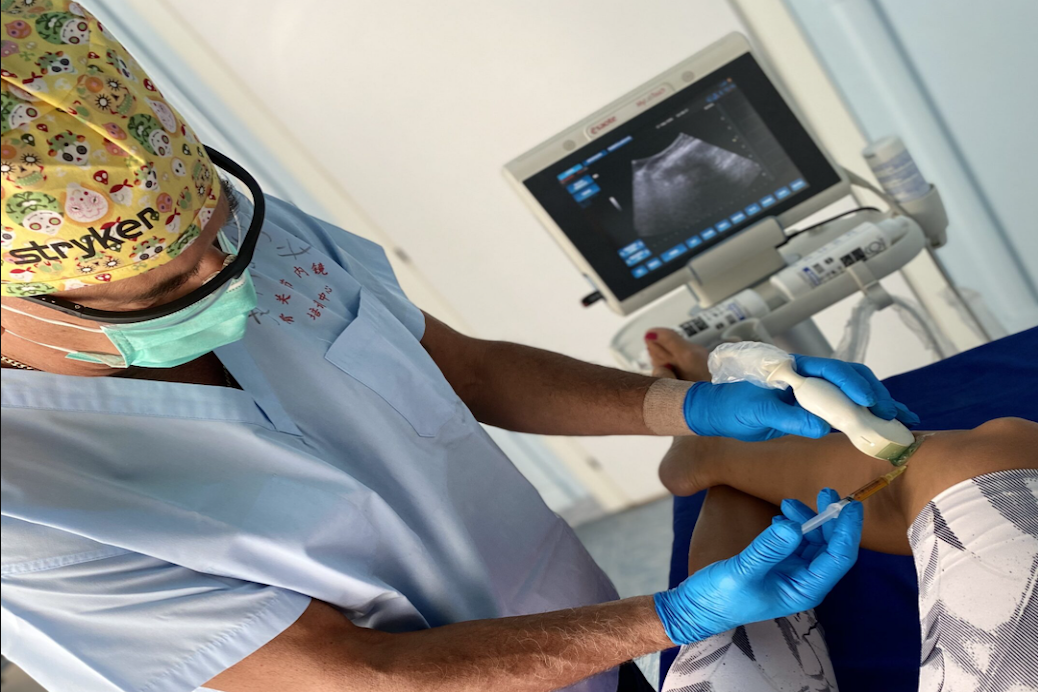
Diagnosis is based on clinical examination, ultrasound and MRI.
CAUSES
Bursitis can be due to repetitive and constant movements in an untrained person (eg long walks or running) or activities that require the use of the goose leg tendons.
TREATMENT
Initially it consists in rest, local ice and NSAIDs, where the disturbances persist it may be indicated to resort to infiltrative therapy with corticosteroids or with biological systems eg. PRP.
A period of abstention from sport and practicing fkt treatments will be useful.
Bone Marrow Edema
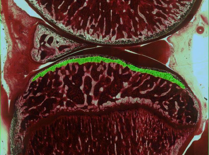
Medullary edema is defined as an imaging alteration that is evident on MRI following trauma or overloading of the subchondral bone: for example, in distorsive knee trauma we often have edema of the condyle and external tibial plateau.
LEARN MORE
SYMPTOMS
The first symptom is pain exacerbated by load on the joint, sometimes redness, heat, and swelling synonymous with an inflammatory picture may appear.
TREATMENT
Initially conservative, consisting of limb unloading, therapy in addition to anabolic drugs and supplements, physical therapies such as magneto therapy.
When it does not resolve and alterations are evident on MRI persisting 3 months later, surgical intervention will be required to stimulate bone healing.
We use biological sub chondroplasty (Osteo-core-plasty technique), a microinvasive method that can be performed on an outpatient or day surgery basis.
RECOVERY
Recovery takes several months especially in injuries found late in life.
Corrective osteotomy
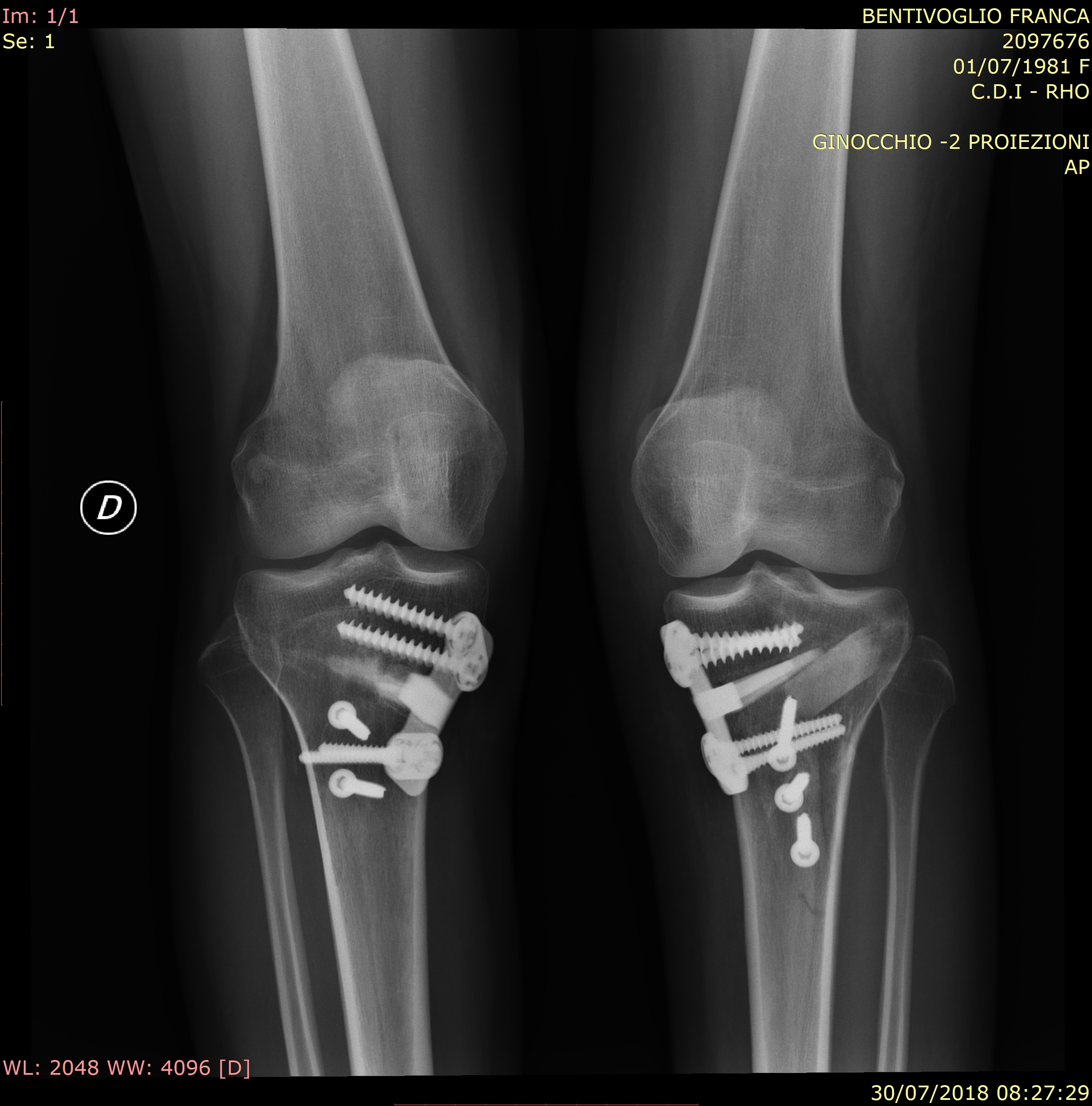
Osteotomy is a surgical procedure that through targeted bone resections with subtraction or addition can’ be able to change the axis of the bone itself.
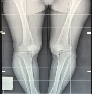
In the knee, this procedure is used when knee arthrosis involves only one side, the medial or the lateral: by limiting the load on the worn compartment it can’ slow the wear and tear of the overloaded part.
By shifting your weight off of the damaged side of the joint, an osteotomy can relieve pain and significantly improve function in your arthritic knee. Knee osteotomy has the goals to transfer weight from the arthritic part of the knee to a healthier area, to correct knee axis malalignment and prolong the life span of the knee joint.
High tibial osteotomy (HTO) has been established as an effective treatment of the varus osteoarthritic knee to decrease the stress on the load-bearing cartilage in the medial compartment.
By preserving knee anatomy, a successful osteotomy may delay the need for a joint replacement for several years.
Another advantage is that there are no restrictions on daily activities and sports participation after an osteotomy.
Due to the overload and wear of the cartilage, the patient complains of localized pain in the worn compartment of the knee, especially at load and in ‘more’ demanding activities.
The osteotomy can be performed with the subtraction technique by resection of a “closing wedge” bone or by “opening” a bone wedge with bone graft in the bone defect created “opening wedge”.
.
Osteotomy is generally associated with other knee “repair” operations such as ligament reconstruction or cartilage transplantation.
If properly performed, the osteotomy surgery unloads the injured part and allows an optimal success of the cartilage transplant, improving the condition and functionality of the patient in a high percentage of cases and delaying the natural evolution of arthrosis and any prosthetic surgery.