


Ankle and Foot
Learn about the most common ankle and foot conditions that we can help you treat
Plantar Fasciitis
There are two plantar arches: one longitudinal, which runs the entire length of the foot and one transverse that runs the width. The arches are made up of ligaments that stabilize the bones. One or both arches can cause pain, but the longitudinal one is more commonly affected.
LEARN MORE
SYMPTOMS
Arch pain is usually caused by overloading during activities such as running, cross- country running, walking, and jumping.
People who have flat feet, or those who have a hyper-pronation problem (when walking, the foot stretches and rotates inwards), are more predisposed to pain in the plantar arch.
Typically, pain occurs slowly.
It can happen suddenly when the ligaments overstretch or tear during strenuous activities, such as shooting or jumping.
DIAGNOSIS
The doctor examines the foot and locates the areas of pain or sensitivity along the arch.
TREATMENT
Ice should be placed on the arch for 20-30 minutes every 3-4 hours for 2-3 days until the pain subsides. The doctor can also recommend taking anti-inflammatory drugs.
Foot support or orthosis may be needed. You can support the arch or insert an insole in the shoe. The doctor may find it necessary to prescribe the use of custom orthotics.
RECOVERY
The purpose of rehabilitation is to return to sport or other usual activities as soon as possible, while ensuring safety. If return to sport or activity is done too early, there is a risk of aggravating the condition, possibly leading to a permanent injury.
Each of us has different recovery times. The speed with which you return to normal activities depends on how the foot heals and not on the days or weeks after the injury.
In general, the longer the interval between the onset of symptoms and the start of therapy, the slower the recovery.
You can go back to practicing your sport safely when, starting from the top to the bottom, you will answer with a “yes” to the following statements:
- The mobility of the affected foot is complete and equal to that of the healthy foot
- You have completely recovered the strength of the affected foot and it is equal to that of the healthy foot
- You can run without pain or limping
- You can take shots without feeling pain or limping
- You are able to flex 45 degrees, first slowly and then at normal speed
- You are able to flex 90 degrees, first slowly and then at normal speed
- You can jump with both feet without feeling pain You can jump with the affected foot only without pain
PREVENTION
It can be prevented by wearing shoes that fit well and have good arch support.
It is useful to stretch the foot and arch before starting sports.
The use of orthotics may be necessary. Some people have to use the insole for the whole day, others only while playing sports.
Tibiotarsal Fracture
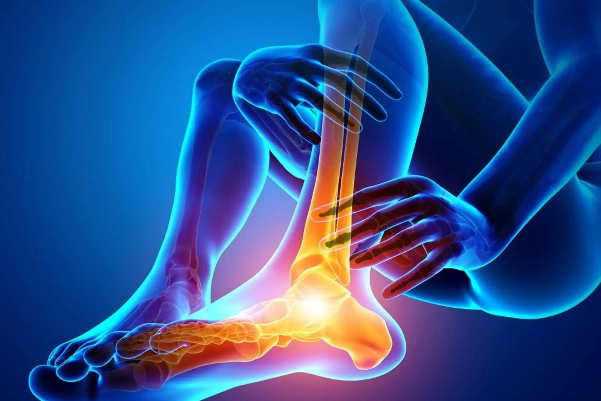
A tibiotarsal joint fracture is a fracture of the tibia and/or fibula near the ankle, causing pain, swelling, bruising, and difficulty bearing weight.
LEARN MORE
CAUSES
A particularly violent trauma can cause fractures in the bone segments that form the ankle.
SYMPTOMS
The symptoms are immediately intense, including pain and swelling, and are accompanied by the inability to walk.
TREATMENT
If the fractures, which usually affect the malleoli, are not very displaced, a knee-high cast is made or a rigid brace is worn, which must be kept for about 35 days.
If, however, the fractured bones have separated too far, surgery is the only way for proper fixation.This involves the use of screws and small metal plates to keep the fragments together.
A period of immobilization and loading protected with crutches will follow.
REHABILITATION
Depending on the injury, the orthopedist will advice when to start rehabilitation. This is essential to retraining incorrect motor patterns learned from prolonged use of the cast.
Your ankle will have completely changed its appearance, it will be difficult to recognize the shape of the malleoli and calf, it will be covered with hard, reddened and dry skin, you will not be able to move it, and you will feel very awkward in walking.
Swimming pool work, especially in the initial stages, is important because it improves venous circulation, retrains a correct motor pattern without difficulty, and strengthens the muscles, while avoiding pain.
Manual therapy is very important and should be accompanied by proprioceptive exercise on foam blocks. Our goals are:
– return to proper ambulation and work activity at 3 weeks after cast removal;
– start treadmill running at 2 months;
– return to sports activity at 3 months after cast removal.
Achilles Tendon Injury
Tendinitis is inflammation of the tendon, which causes pain in the back of the leg and heel. The inflammation causes pain in the back of the leg, at the level of the heel.
LEARN MORE
INJURY
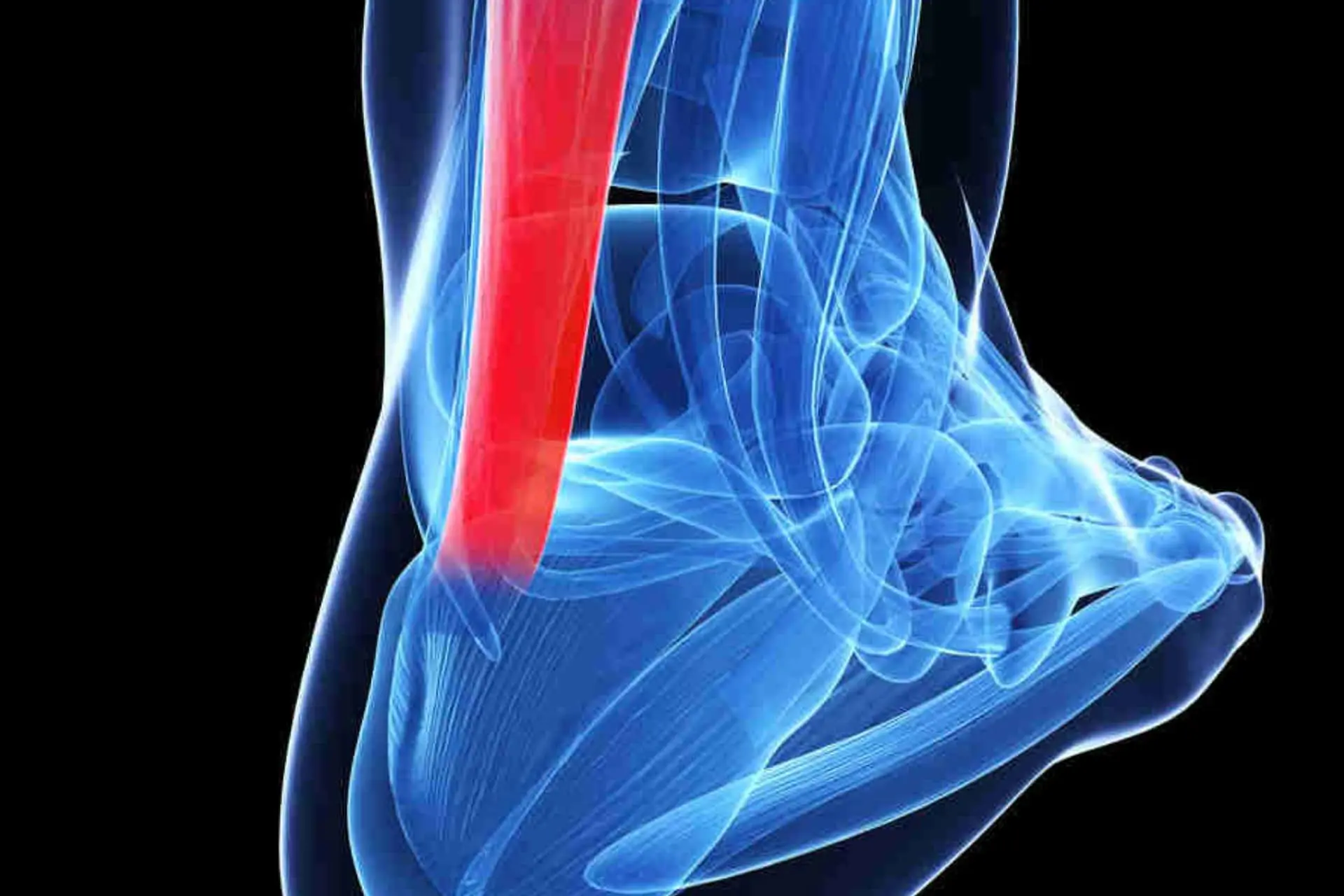
The Achilles tendon is a band of tissue that connects the heel to the calf muscles.
Tendon injuries can be inflammations or even tears.
Tendinitis is inflammation of the tendon, which causes pain in the back of the leg and heel. The inflammation causes pain in the back of the leg, at the heel level.
Tears can be a full or partial rupture of the tendon and it also causes pain in the heel.
CAUSES
Tendinitis can be caused by:
- Excessive use of the Achilles tendon.
- A short calf muscle.
- A short Achilles tendon.
- Excessive efforts uphill.
- Increase in training intensity, sometimes on rough terrain, and use of poorly “cushioned” running shoes.
- Hyper-pronation: during walking or running the foot rotates inwards more than normal.
- Use of high-heeled shoes at work and flat shoes for sports.
- The Achilles tendon can rupture during exercises that involve rapid efforts. For example, by jumping or doing sprints. It mainly affects jumpers, runners, football and tennis players, who usually employ sharp muscle contraction in their movements.
SYMPTOMS
Tendinitis causes pain and sometimes swelling in the Achilles tendon. The tendon is painful and swollen. Pain is felt when standing on tiptoes and when stretching the tendon.
Ankle mobility is limited. When the tendon breaks, a snap is heard. When the tear is complete, you are no longer able to lift the heel or load on the toes.
Acute and sudden pain felt at the level of the Achilles tendon, functional inability, and palpating a gap in the tendon are basis of diagnosis and confirmed by ultrasound investigation.
DIAGNOSIS
The doctor examines the leg, checking for painful areas and for swelling. Observe your foot while walking or running to see if there is hyper-pronation.
TREATMENT
Put ice on the Achilles tendon for 20-30 minutes every 3-4 hours for 2 or 3 days until the pain disappears.
When lying down, the lower leg should be raised by placing a pillow underneath.
Take the prescribed anti-inflammatory medicines.
If a cushion is prescribed to be placed in the shoe, under the heel, it must also be used after healing of the tendon. The pad prevents excessive stretching of the Achilles tendon.
During the rehabilitation phase, another sport, or activity, that does not aggravate the condition must be practiced. For example, you can swim instead of running.
Tendon stretching and strengthening exercises recommended by the therapist should be performed.
If it is a problem of hyper-pronation of the foot, the doctor will prescribe a customized insole, called orthotics, to be inserted in the footwear in order to make the foot more stable.
In cases of severe tendinitis, the foot should be immobilized for 6-10 weeks.
Surgical Treatment
By different types of suture of the tendon, called TENORRHAPHY; sometimes, a reinforcement is made using the tendon of the footbed. The intervention is usually followed by immobilization with a rigid brace for 3 or 4 weeks, then replaced by a Cam Walker with which you begin to walk gradually.
Rehabilitation Treatment
Rehab lasts about 12 weeks and includes massages on the scar and calf muscles, physical therapies for pain and swelling, exercises for the joint and for the restoration of strength and correct walking and running.
After about 3 months, it is possible to carry out an ISOKINETIC TEST to measure the muscle strength of the leg. Based on the result, the therapeutic protocol is adapted to bring the athlete back to the sports field where, together with the coach, he re-trains coordination of the specific technical movements of the sport.
RECOVERY
The purpose of rehabilitation is to return to sports or other usual activities as soon as possible, but in complete safety.
If it is too early, there is a risk of aggravating the conditiong and this could lead to a permanent injury.
Everyone has different recovery times.
How quickly you return to normal activities depends on how the ankle heals, not on the days or weeks that have passed since the injury. In general, the longer the interval between the onset of symptoms and the start of therapy, the slower the recovery.
You can go back to practicing your sport safely when, starting from the top to the bottom, you will answer with a “yes” to the following statements:
- The mobility of the affeced leg is equal to that of the healthy leg.
- You can you stretch the affected leg like the healthy one.
- You can run straight without pain or limping
- You can take shots without feeling pain or limping
- You are able to flex your foot at 45 degrees, first slowly and then at normal speed.
- You are able to flex your foot at 90 degrees, first slowly and then at normal speed.
- You can jump with both legs without feeling pain. You can jump with the affected leg only without pain.
PREVENTION
The best prevention is to stretch the calf and Achilles tendon muscles before doing sports.
If you have a short Achilles tendon and short calf muscles, you need to stretch 2 times a day, even if you don’t play sports.
If you have a predisposition for tendinitis, avoid running uphill for a long time.
Metatarsalgia
Metatarsalgia is the pain that is felt in the long bones of the foot, especially at the level of the head, or toe.
LEARN MORE
CAUSES
Metatarsalgia typically arises due to excess loading such, as running, jumping, or walking.
It can also arise simply because of a new pair of shoes, especially if with a high heel.
There are people in whom the tips of some metatarsal bones are more inclined downward than the norm, so they are more likely to cause pain.
SYMPTOMS
Mid-foot pain, especially “over” the bones.
DIAGNOSIS
The specialist examines the foot and orders an x-ray of the foot to check that there are no fractures. In the event that no fractures are identified, it is metatarsalgia.
TREATMENT
It can be treated with anti-inflammatory drugs. The specialist can recommend the use of an orthotic support to be placed under the painful metatarsal area.
Custom arch supports are often prescribed.
During rehabilitation, it is necessary to replace sport and usual activities with others that do not aggravate the condition.
For example, swimming or cycling instead of running or walking.
RECOVERY
The purpose of rehabilitation is to return to sports or other activities as soon as possible, but in complete safety. If it is too early, there is a risk of worsening the situation and this could lead to a permanent injury.
Each of us has different recovery times. Returning to your sport, or activity, will depend on how quickly your foot heals, not on the number of days or weeks after the injury.
In general, the longer the interval between the onset of symptoms and the start of therapy, the slower the recovery.
You can go back to practicing your sport safely when, starting from the top to the bottom, you will answer with a “yes” to the following statements:
- Do you have complete mobility of the affected foot and is it equal to that of healthy foot?
- Can you jump with both feet without feeling pain?
- Can you jump with the affected foot only without pain?
PREVENTION
The best prevention is to wear comfortable shoes that fit well.
Morton's Neuroma
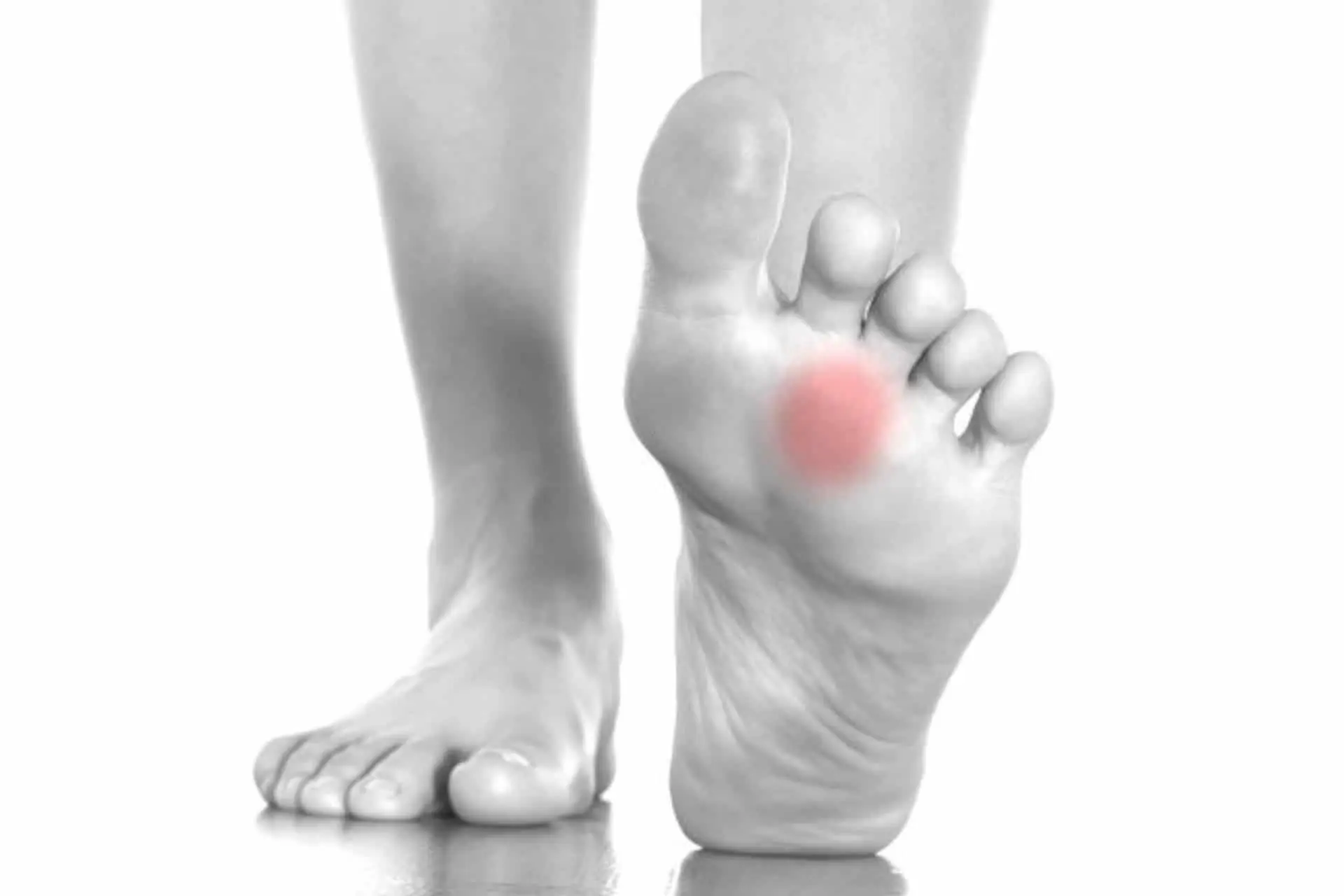
There are nerves between the bones of the foot and along the toes. A neuroma is a benign (non-cancerous) tumor of the nervous tissue. Morton’s Neuroma is most commonly located between the bones of the second and third toes or between the third and fourth.
LEARN MORE
CAUSES
A neuroma can form due to excessive running or walking, or as a result of the use of tight or high-heeled shoes. Sometimes it forms in the absence of obvious causes. The pain worsens when walking on hard surfaces or wearing footwear which are too tight.
SYMPTOMS
The foot is sore. The pain usually intensifies when the toes are pushed upwards. The affected area may be itchy or be numb. It occurs most often between the second and third or third and fourth toes.
DIAGNOSIS
The doctor examines the foot, assesses the symptoms, and the location of the pain. The doctor examines the foot, assesses the symptoms, and the location of the pain.
TREATMENT
In order of treatment:
- Wear shoes that fit properly.
- Take anti-inflammatory drugs.
- Wear an orthotic support under one of the bones of the foot or use a customized arch support.
In case these suggested treatments fail, cortisone injections can be tried. Surgery may be needed to remove the neuroma. Surgery may be needed to remove the neuroma.
PREVENTION
To date, there is not a therapy for preventing its formation. However, the use of properly fitting shoes and appropriate insoles helps to reduce pain.
Athlete's Foot
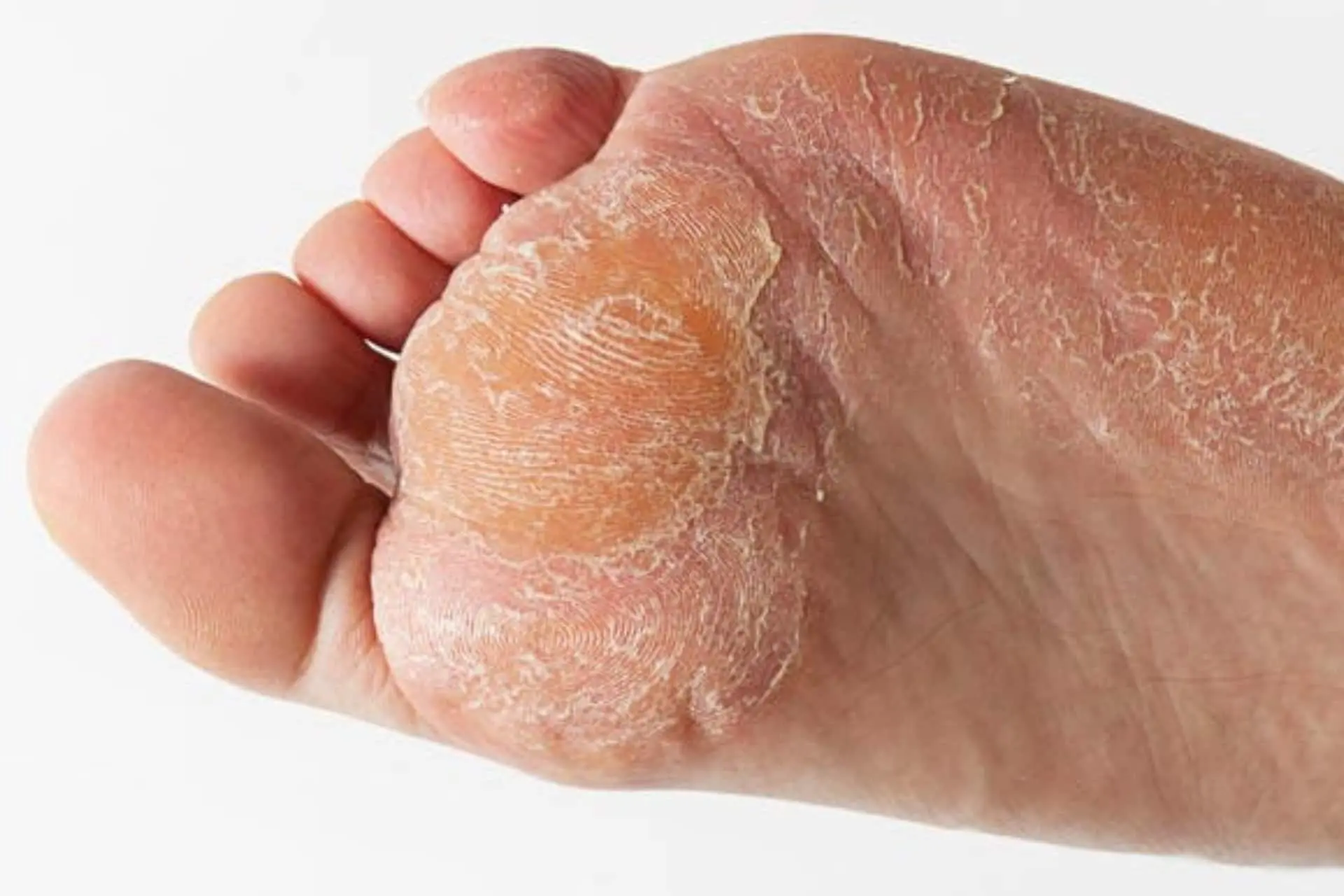
Athlete’s foot is a common skin problem and is caused by a fungus. In general, it initially affects the skin between the toes that becomes scaly and itchy.
LEARN MORE
INJURY
Over time, it can lead to the laceration of the skin and pain.
Another term for athlete’s foot is tinea pedis (ringworm of the foot).
CAUSES
The fungus that causes athlete’s foot is found everywhere. Generally, it is found on the floors of showers, changing rooms, and in the gym.
The fungus proliferates more easily on sweaty, damp and, badly dried feet (especially if you then wear shoes and socks with little aeration).
SYMPTOMS
Symptoms are:
- Itching
- Chapped and scaly skin. Usually between the last two toes
- Pain
- Blisters (sometimes)
DIAGNOSIS
In general, the doctor diagnoses athlete’s foot by examining the skin. Sometimes, he can take a sample of skin to analyze the fungus.
In the event that a bacterial infection is also suspected, bacteriological tests will be carried out.
TREATMENT
The infection can resolve itself without being treated, but in most cases, medicines are used on the skin.
If the infection is severe and extensive, it may be appropriate to take oral medications.
RECOVERY
The acute infection phase lasts from 1 to 10 days. Chronic infection can persist for months or years. If a serious infection is not treated, it can lead to a serious bacterial infection.
The infection can also affect the nails, and in this case, treatment will be more difficult.
PREVENTION
Follow these guidelines:
- Wear cotton socks when exercising
- Change your socks daily
- Wear sandals or shoes with ventilation holes or porous material (a natural material such as canvas or leather, rather than synthetic materials)
- Allow shoes to air out when not in use
- Wear sandals when using public showers
- Dry your feet very well, especially between your toes
- Apply an anti-fungal powder to the parts at risk
- Disinfect the floor of the shower and dressing room
Achilles Tendinopathy
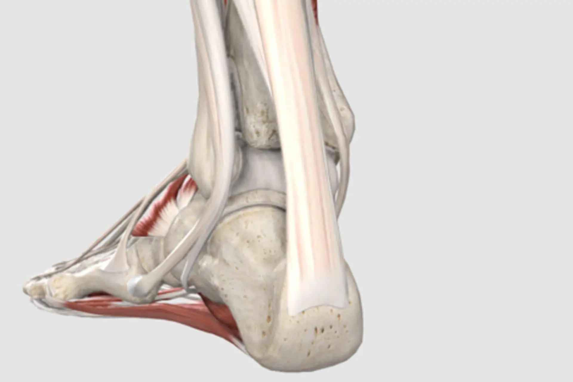
Inflammation of the Achilles tendon-tendonitis-is a particularly common and feared occurrence in sports.
LEARN MORE
INJURY
Ground quality, use of ill-fitting shoes, inadequate warm-up, excessive loads, poor coordination, and anatomic or physiologic problems, can generate repeated microtraumas and high-intensity functional stresses.
Stress on the tendon for particularly long periods of time can cause inflammatory and degenerative lesions.
SYMPTOMS
Symptoms can begin gradually, but in severe cases, they can affect sports performance and ability to play.
DIAGNOSIS
It is characterized by pain, swelling, redness of the skin, and is confirmed by ultrasound, which clarifies the location, degree, and extent of the lesion.
TREATMENT
It is essential to start rehabilitation treatment from the onset of symptoms. The treatment of a tendinitis is always very delicate, and the chances of success depend on the severity of the pathology and the time of onset of the symptoms.
In our experience, based on the treatment of many athletes, including professionals, we have learned that it is essential to address even the slightest signs of disease, setting up a personalized rehabilitation treatment early, which in principle consists of:
- Active rest: reducing, varying, or suspending sports activities according to the gravity of the situation;
- Drug therapy and physical therapy;
- Shockwave therapy: we have recently introduced this therapeutic method with excellent results;
- Manual therapy: it offers a significant contribution in the hands of a good therapist;
- Stretching exercises, muscle strengthening, proprioceptive re-education;
- Baropodometric evaluation with consequent use of corrective insoles or bilateral raises to unload the tendon;
- Evaluation of the chiropractor to resolve joint blockages of the foot, often present in chronic injuries;
- PRP treatment
Ankle Sprain Trauma
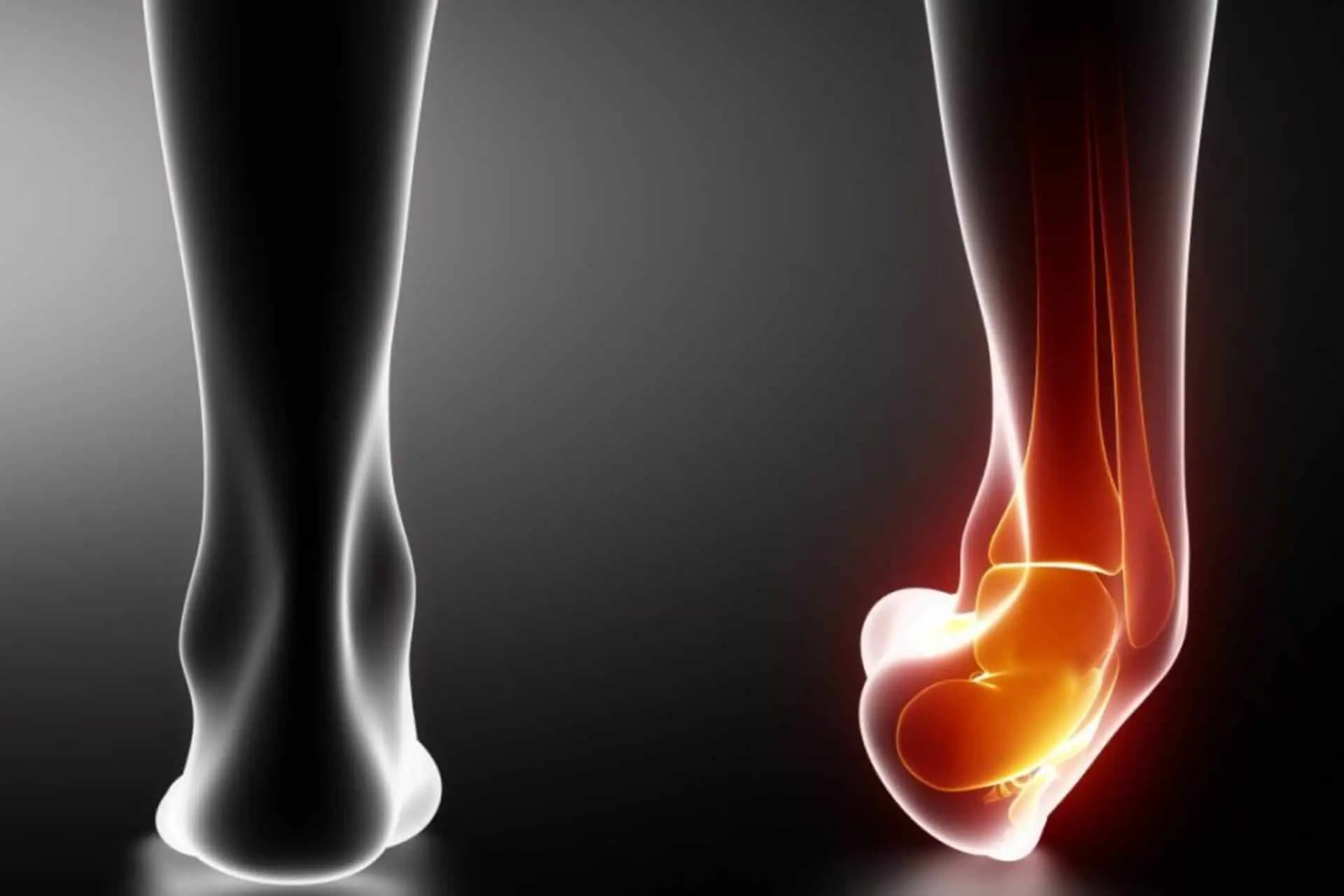
Ankle sprains are ligament injuries caused by excessive twisting or rotation of the ankle.
LEARN MORE
We divide the treatment into 5 phases:
First phase: lasts about 10 days, we protect the joint with a brace and possibly also with the use of crutches, we try to minimize the inflammatory picture and bruising by using ice and physical therapies, we aim to obtain an accurate diagnosis.
Second phase: Dorsi and plantar flexion is restored using a swimming pool with high water and unstable surfaces to recover the ability to react to external insults, even more important in a joint such as the ankle. We gradually increase the intensity of the exercises and the load.
Third phase: dedicated to the recovery of correct walking and articulation.
Fourth phase: we recover muscle strength by seeking a good balance between the medial and lateral muscles of the ankle, which is essential to restore at least the dynamic part of stability, we also refine the proprioceptive work with increasingly complex paths and with leaps on the trampoline. Before moving on to the last phase, in which the athletic movement is reworked, we measure the recovered strength, comparing it with the healthy limb, through the ISOKINETIC TEST.
Fifth phase: collaboration with the coach in the sports field is essential, in fact our objectives are:
– recovery of coordination and dexterity present before the trauma;
– prevention of new traumas;
– avoiding chronic instabilities typical of many athletes, but especially basketball players. In this case, the RECONDITIONING of the ankle.
Surgical treatment of external ligaments can be indicated in very severe chronic instability pictures. The intervention is followed by approximately 35 days of immobilization and subsequent re-education.
Sport is expected to resume after 4 months.
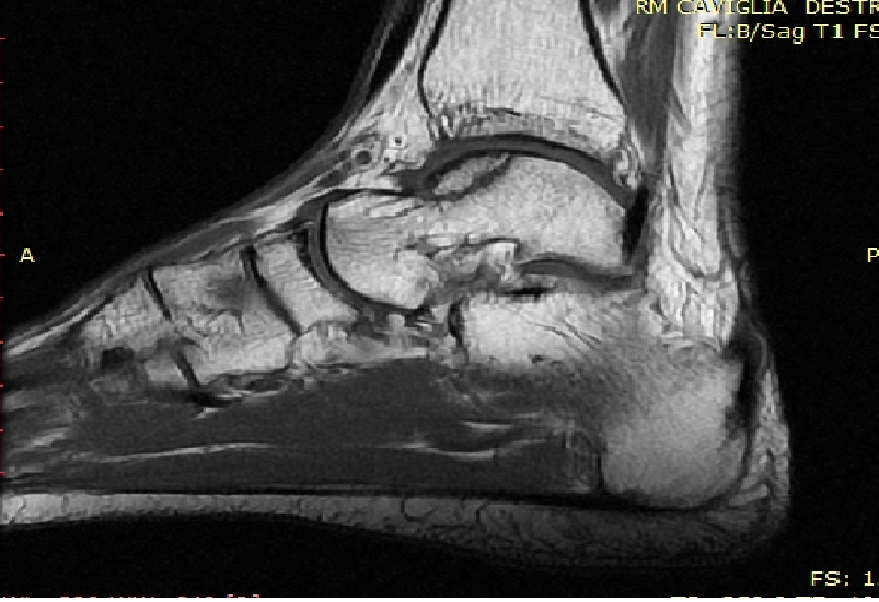
Osteochondral Injury Of The Talus
An osteochondral lesion of the talus is damage to the underlying cartilage and bone in the ankle joint.
LEARN MORE
Osteochondral lesions of the talus are an important cause of ankle pain and disability. There is a wide variability in the literature reporting about the incidence of such injuries due to the high frequency of sprained ankles and a scarcity of adequate diagnostic investigations. An osteochondral lesion of the talus can be the cause of progressive chondral damage that can lead to osteoarthritis and profound disability. Whenever we see persistent pain after an ankle sprain, we should think about possible osteochondral damage.
ETIOLOGY
There are multiple causes that can lead to an osteochondral lesion of the talus: trauma and ischemia are often reported as possible etiologies, although other possible associations with endocrine disorders, including genetically transmitted ones in some cases, have been identified. However, the percentage of association of osteochondritis dissecans of the talus with previous traumatic episodes is not clear. However, it has been demonstrated as a possible cause in some studies on cadavers. It is important to consider that the association of an osteochondral lesion with a previous trauma is generally linked to the patient’s story and can be problematic given the high incidence of trauma to the ankle.
CLASIFICATION
Ferkel and Sgaglione: Classification based on CT images
|
Stage I |
Cystic lesion within dome of talus, intact roof on all views |
|
Stage IIA |
Cystic lesion with communication to talar dome surface |
|
Stage IIB |
Open articular surface lesion with overlying non-displaced fragment |
|
Stage III |
Non-displaced lesion with lucency |
|
Stage IV |
Displaced fragment |
Today, however, we must consider that MRI offers higher quality and precision images.
Mintz et al. MRI-based Classification
|
Stage 0 |
Normal |
|
Stage 1 |
Hyperintense cartilage surface, morphologically intact |
|
Stage 2 |
Fibrillation or fissures not extending to bone |
|
Stage 3 |
Flap present or bone exposed |
|
Stage 4 |
Loose, non-displaced fragment |
|
Stage 5 |
Displaced fragment |
TREATMENT
A BIOLOGICAL problem must be treated with a BIOLOGICAL treatment.
Only a few cases in young patients with acute lesions can procedures be made to reattach the osteochondral fragment.
Cartilage lesions can be treated by shaving, microfractures, cartilage transplantation or grafting with mesenchymal cells. If the lesion is subchondral, we recommend the Osteo-Core-Plasty technique that we have created.
OSTEO-CORE-PLASTY TECHNIQUE
We begin with the removal of the bone marrow aspirate from the iliac crest using a dedicated kit from Aspire. Using a special introducer, the cell-rich medullary space is entered up to the desired depth. A small quantity of marrow is then removed at different depths by gradually retracting the cannula in order to obtain the maximum possible quantity of marrow cells. Once the desired quantity has been obtained, it can be applied using an arthroscopic technique under amplioscopic control.
This video illustrates the treatment of a large osteochondral lesion treated arthroscopically.

