


Elbow and Wrist
Learn about the most common elbow and wrist conditions that we can help you treat
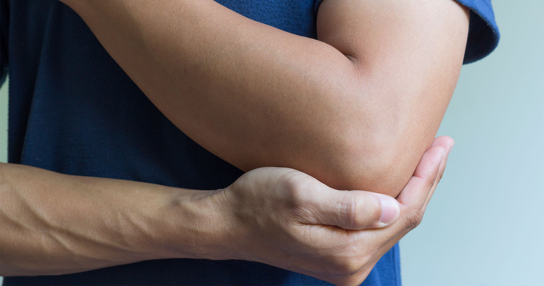
Epicondylitis
Epicondylitis consists of tendon damage (tendinopathy) of the muscles that insert on the epicondyle of the elbow (laterally) that move the forearm in supination, along with wrist and finger extension.
LEARN MORE
CAUSES
The most common cause is chronic micro-traumatic stress as a result of repeated efforts involving the elbow muscles (e.g. tennis, manual work like an electrician, etc.). However, it can also present acutely, following an isolated stress or trauma.
SYMPTOMS
Pain localized at the insertion of the tendons on the lateral epicondyle of the elbow with pressure and/or maneuvers of the carpus and fingers such as extension, pronation, or supination (e.g. pouring from a bottle, opening a handle, using a screwdriver, tightening the hand, etc.)
TREATMENT
Acute phase (7-20 days):
- Functional rest (avoid maneuvers that activate the affected muscles and cause pain).
- Local ice (20 min 2-3 times a day, or alternatively massage with an ice cube for 5 minutes).
- Nonsteroidal anti-inflammatory drugs (NSAIDs) according to medical prescription.
- Physical therapy (laser therapy, iontophoresis, ultrasound, TENS) according to medical prescription.
Subacute phase (20-60 days):
- Forearm massage therapy.
- Stretching of epicondyloid, epitroclear, and shoulder-humeral girdle muscles.
- Gradual active exercise against resistance (elastic, weight), mainly eccentric for at least 6 weeks.
- Possible continuation of physical therapy.
- Use of epicondylitis brace for activities that can strain the elbow.
- Any applications of extracorporeal shock waves (according to medical prescription).
- Local infiltrative treatment with autologous platelet concentrate (P.R.P.) rich in growth factors that promote tissue regeneration and real healing. The treatment is performed with the use of an ultrasound machine to make the infiltration more precise.
PRECAUTION
- Avoid activities that provoke and maintain pain, especially in the acute phase.
- Avoid corticosteroid infiltrations that only temporarily relieve the symptoms.
- Do not underestimate the pathology, treat it early. As pathology advances, treatment becomes slower and more difficult.
RECOVERY
Recovery occurs after resolution of the painful symptoms and functional re-education of the elbow with recovery of adequate tone and muscle flexibility.
The resumption of sports activities must be carried out with medical consent and with the possible use of a specific brace.
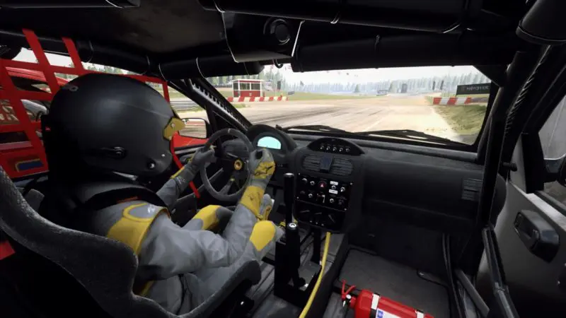
Driver's Elbow
Driver’s Elbow occurs due to keeping the elbow flexed and resting on the upper edge of the door (the typical elbow outside the window).
LEARN MORE
DIAGNOSIS
This pathology can often be confused with the more common, “golfer’s elbow” or cervical radiculopathy. Therefore, it is always recommended to consult a specialist who, through careful physical examination and the aid of instrumental investigations, will be able to formulate a correct diagnosis. Diagnosis is important because this pathology can often be confused with the more common, “golfer’s elbow” or cervical radiculopathy.
SYMPTOMS
Driver’s elbow will commonly present as pain, generally dull and continuous, which can be associated with paresthesia, tingling, and in the most severe cases, motor deficit up to the contracture in flexion of the fingers innervated by the ulnar nerve (fourth and fifth finger in particular).
TREATMENT
The therapy is divided between the simple intake of anti-inflammatory drugs, possible PRP infiltrations, as well as physical therapies, up to, in the most complicated cases, surgical decompression of the ulnar nerve.
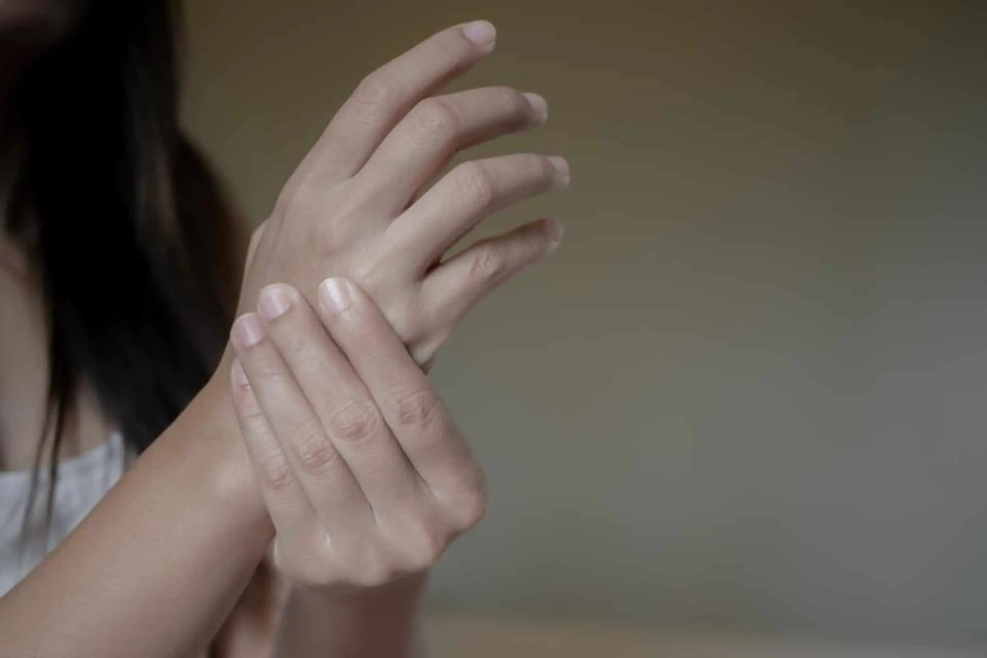
Carpal Tunnel Syndrome
Carpal tunnel syndrome is a fairly common disorder that affects the functionality of the hand. This condition appears when the median nerve is compressed as it passes through the Carpal tunnel.
LEARN MORE
CAUSES
- Dislocations or fractures: as a result of dislocations or fractures, a bone protrusion forms in the canal which then creates additional pressure and compresses the median nerve
- Wear: caused by chronic, repetitive hand movements
- Fluid retention: this condition appears more frequently during pregnancy, and symptoms can disappear with childbirth.
SYMPTOMS
Carpal tunnel syndrome presents with a reduction in sensitivity to touch, with pain, and tingling, which in particular appear at night. It is a disorder not to be overlooked, as it can progressively degenerate.
PREVENT THE SYNDROME
- Keeping the wrist in a neutral position, i.e. avoiding over flexion, extension or rotation for an extended period of time.
- Avoiding repetitive movements, trying not to hold an object in the same way for long periods of time
- Resting your hands with short breaks
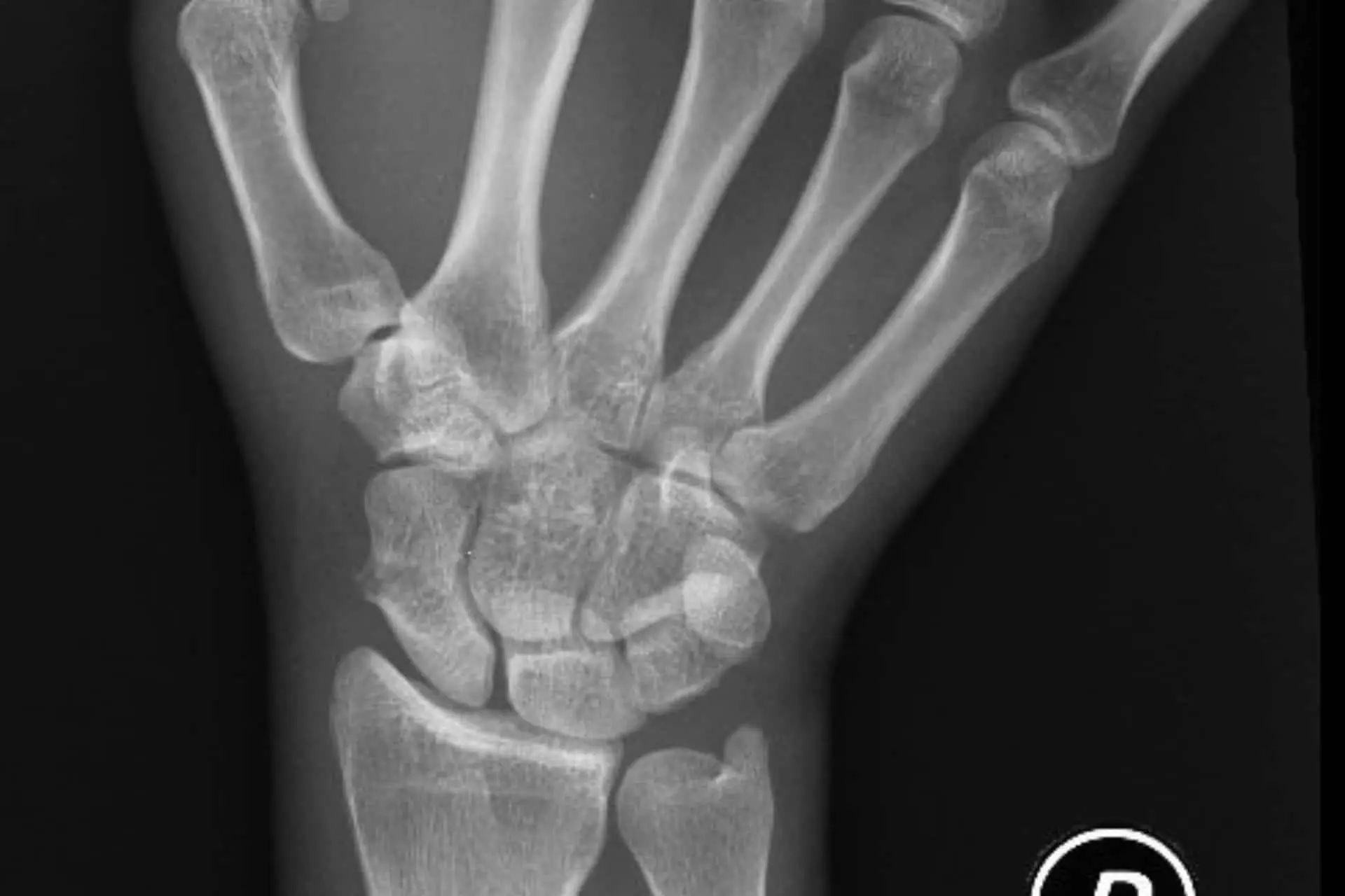
Avascular Necrosis Of The Scaphoid
This occurs when impaired blood flow leads to bone death, causing wrist pain
LEARN MORE
INJURY
Carpal tunnel syndrome is a fairly common disorder that affects the functionality of the hand.
The scaphoid bone is extremely susceptible to this complication because the vascularization of the bone is terminal, and these small vessels are often damaged during fracture of the scaphoid.
This results in the absence of blood supply to nourish the bone which consequently leads to necrosis.
CAUSES
The most common causes are fractures during hyperextension of the wrist or following a road accident, with a compression trauma generated by the reaction force that is applied to the steering wheel by the forearm.
There is also an idiopathic form of this pathology, known as “Preiser disease”.
SYMPTOMS
AVNS most commonly presents as pain in the wrist, which radiates from the base of the thumb along the radial side of the forearm.
This pain can be exacerbated during movements of the wrist or thumb, both active and passive.
DIAGNOSIS
Diagnosis requires imaging techniques such as X-ray, CT, or MRI.
TREATMENT
Treatment can be pharmacological, initially, accompanied by temporary immobilization of the wrist.
Local ice application can help ease pain. The long-term treatment of choice remains to be surgery. Therefore, consultation of a specialist is absolutely recommended.
Pump Arm Syndrome
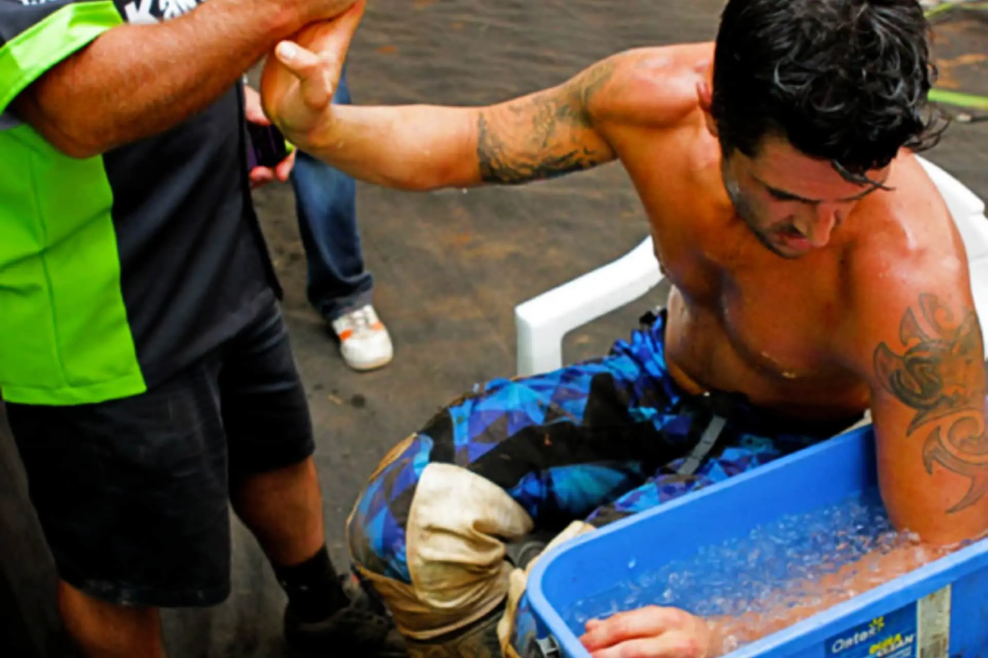
Pump arm syndrome is a condition caused by prolonged use of vibrating tools, which causes pain in the hand.
LEARN MORE
PUMP ARM SYNDROME
The “Pump Arm Syndrome” or the painful contracture of the forearm in Motocross, is a difficult problem to solve.
The painful contracture of the forearm is a frequent and very annoying problem for the motocross rider. It affects all levels of riders from the Sunday enthusiast to the professional driver and is sometimes so debilitating that it forces the rider to abandon the sport.
It can be very dangerous as it causes a decrease in the ability to control the motorcycle and can cause serious accidents.
It is a problem frequently encountered in motocross and less in other popular sports. Few studies have been conducted on the subject since no motorcycle manufacturer or sports federation has ever invested money by supporting foundations or research institutes to investigate the mechanisms that determine this problem.
It is not uncommon, in fact, for the motocross rider to go to a sports or orthopedic doctor complaining of the “pump arm syndrome” and the doctor does not even know of the existence of this pathology.
As motocross readers know, for over 30 years, I have been dealing with off-road vehicles, first as a driver and then as a sports doctor: I will try to shed some light on the problem.
First of all, I want to underline a curious aspect. I did a research of the current literature and this pathology is very poorly known and there are few results reported after surgical treatment. However, by simply attending motocross races, even at a lower level, everyone knows what Pump Arm Syndrome is. Riders can remember suffering from painful contracture of the forearm and recount their experiences. The most varied attempts of treatment range from massages with grandmother’s cream to complicated surgical interventions which are only performed strictly in Belgium, one of the hotspots of motocross!
The problem is complex, and we must start from the beginning. What are the causes that can determine the pain in the forearm in the motocross rider?
A plethora of factors may be involved, but the most common manifestation is the “chronic compartment syndrome of the forearm.” After a few laps of the race, a painful contracture of the entire forearm with pain and numbness of the fingers results in a partial or total impairment of the ability to close one’s hand and hold the handlebar.
It is important to distinguish between chronic (most common) and acute (fortunately rare) onsets of Pump Arm Syndrome. In chronic cases, where the rider complains of pain and a contracture, the rider must stop riding and the disorder should gradually decrease with rest. An acute compartment syndrome in which the rider can sustain a fracture or tear a muscle during a fall, the case is much more serious. In this case, there is intrafascial bleeding and the subject will experience rapid forearm swelling and sudden loss of the sensitivity in the fingers.This requires immediate hospitalization in an emergency room!
The rider must try to passively move the fingers and go immediately to a center specialized in these pathologies.
Since it has now been understood that the problem is related to blood circulation in the forearm, possible solutions can be analyzed:
NON-SURGICAL THERAPY
Over the years we have developed a series of therapies-exercises that can solve most cases.
We start with training, eliminating weight-bearing exercises that hypertrophy the forearm muscles and can worsen this syndrome. We provide a list of useful activities for the pilot forearm muscles. In established pathologies we use infiltration with a particular type of PRP into the forearm bands.
SURGICAL THERAPY: we are often used to considering surgery as a definitive solution to the problem; we must not forget the possible failures and complications of such surgery.
My “Anglo-Saxon” mentality, scientifically speaking, has led me to always explain to patients what are the results reported in the literature about the surgery to be performed and what is my experience.
The “fasciotomy” surgery, ie the opening by incision of the bands that surround the muscles of the forearm, is normally performed in emergency situations for acute compartment syndrome and does not present particular difficulties.
Having said that, it is important to know that there are few well-documented studies about the effective utility in the “Pump Arm Syndrome”. The result is, on average, positive in about 50% of, but some athletes have shown a worsening after this surgery due to the formation of scar tissue.
The operation can be performed in day-hospital with regional local anesthesia and the return to sport usually takes place after a month.
Ulnar Tunnel Syndrome
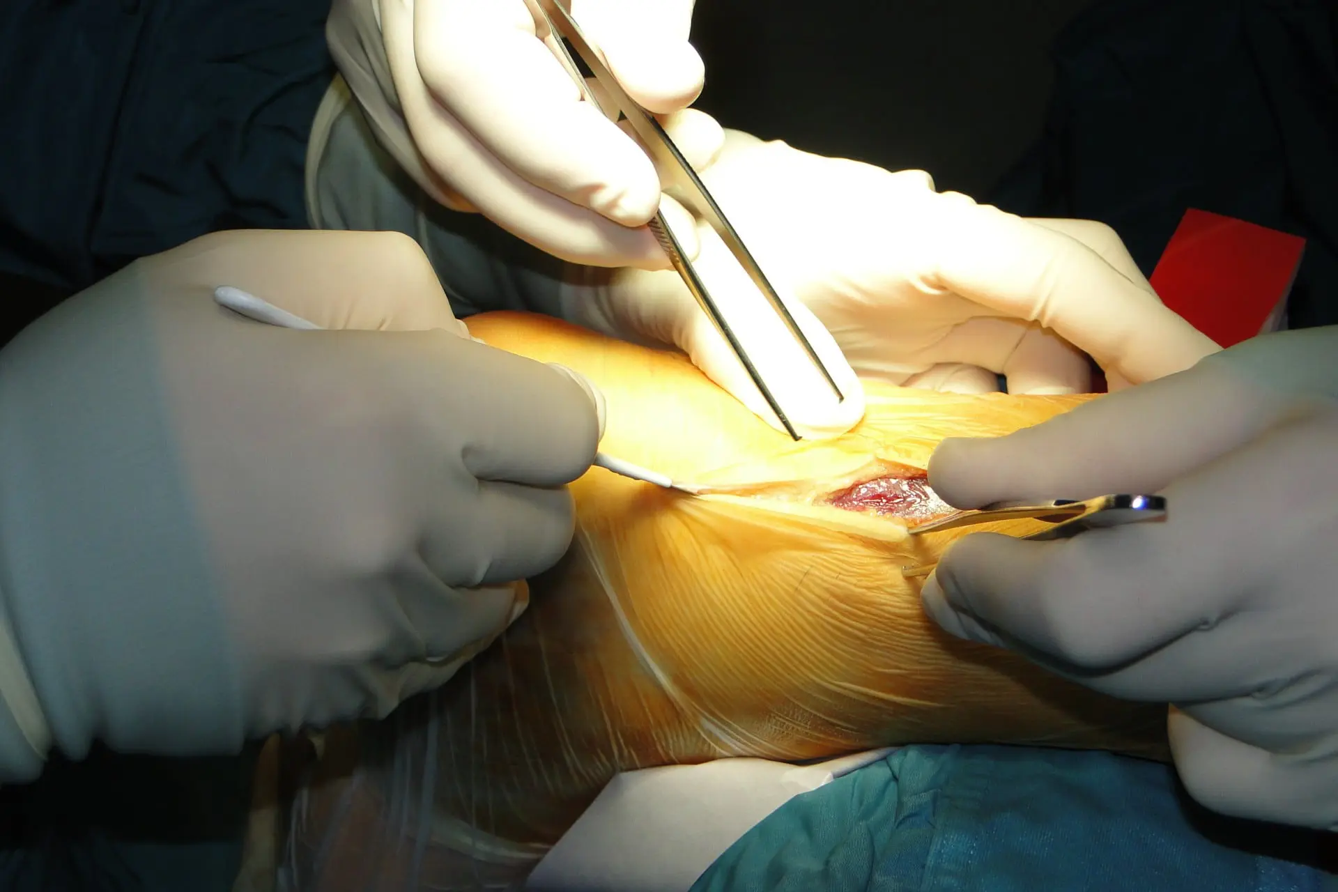
It is a neuropathy from entrapment of the ulnar nerve at its point of passage from the forearm to the hand, called Guyon’s canal. It is characterized by pain, numbness and paresthesia of the wrist, which radiates to the ulnar side of the palm and the last two fingers.
LEARN MORE
CAUSES
It is a syndrome with higher incidence in the female sex, and the clinical onset usually occurs as a result of repetitive wrist movements or after direct trauma to the area of the palm of the hand called the “hypothenar eminence,” located at the fifth finger of the hand.
SYMPTOMS
The pain is of a burning type, present most at night, with paresthesia generally described as “needles that pierce.” Paresthesias are generally motor, although sometimes they can be mixed sensory-motor.
DIAGNOSIS
Diagnosis is confirmed with a postive Tinel’s sign, a test conducted by the specialist and signaling a possible weakness of the intrinsic muscles of the hand.
TREATMENT
Treatment can be started with anti-inflammatory drugs, associated with rest, but it is highly recommended to consult a specialist because it may be necessary to resort to infiltrations to reduce pain or to intervene surgically.

Anterior Interosseous Syndrome
It is an uncommon cause of forearm and wrist pain. The onset of symptoms usually results from acute trauma to the forearm or repeated movements of the forearm and elbow.
LEARN MORE
CAUSES
Generally, this syndrome is due to a compression of the median nerve at the belly of the round pronator muscle and the superficial flexor of the fingers.
SYMPTOMS
Clinically, this syndrome manifests as acute pain in the proximal forearm, with possible involvement of the wrist.
The most frequent clinical signs are inability to flex the distal phalanges of the index and middle fingers and the proximal phalanx of the thumb.
Tinel’s sign, a clinical test usually evaluated by the specialist, may be positive.
TREATMENT
Treatment may require anti-inflammatory drugs or other drugs that must be prescribed only after a specialist visit. Infiltrations may be prescribed to relieve pain, and if symptoms persist, surgery may be required.

Biceps Brachii Tendinitis
It is an increasingly common clinical syndrome, especially since the number of people getting into fitness has increased. The portion of the biceps most prone to inflammation is the distal tendon, which fits over the radius.
LEARN MORE
CAUSES
Generally, the most common causes are microtraumatic injuries due to repetitive movements, especially during the performance of certain exercises in the course of physical activity.
Chronic inflammation can also lead to the development of calcifications at the level of the tendon, which will make treatment more difficult. Repeated trauma can lead to tendon rupture.
SYMPTOMS
Symptoms appear mostly acutely, for example playing tennis, by lifting a weight or performing exercises in the gym that involve the use of the biceps. The pain is constant and severe, localized in the crease of the elbow and can affect the quality of sleep.
DIAGNOSIS
Physical examination by a clinician is important to evaluate the function of the biceps. Additional imaging, such as X-rays or MRI, may be necessary.
TREATMENT
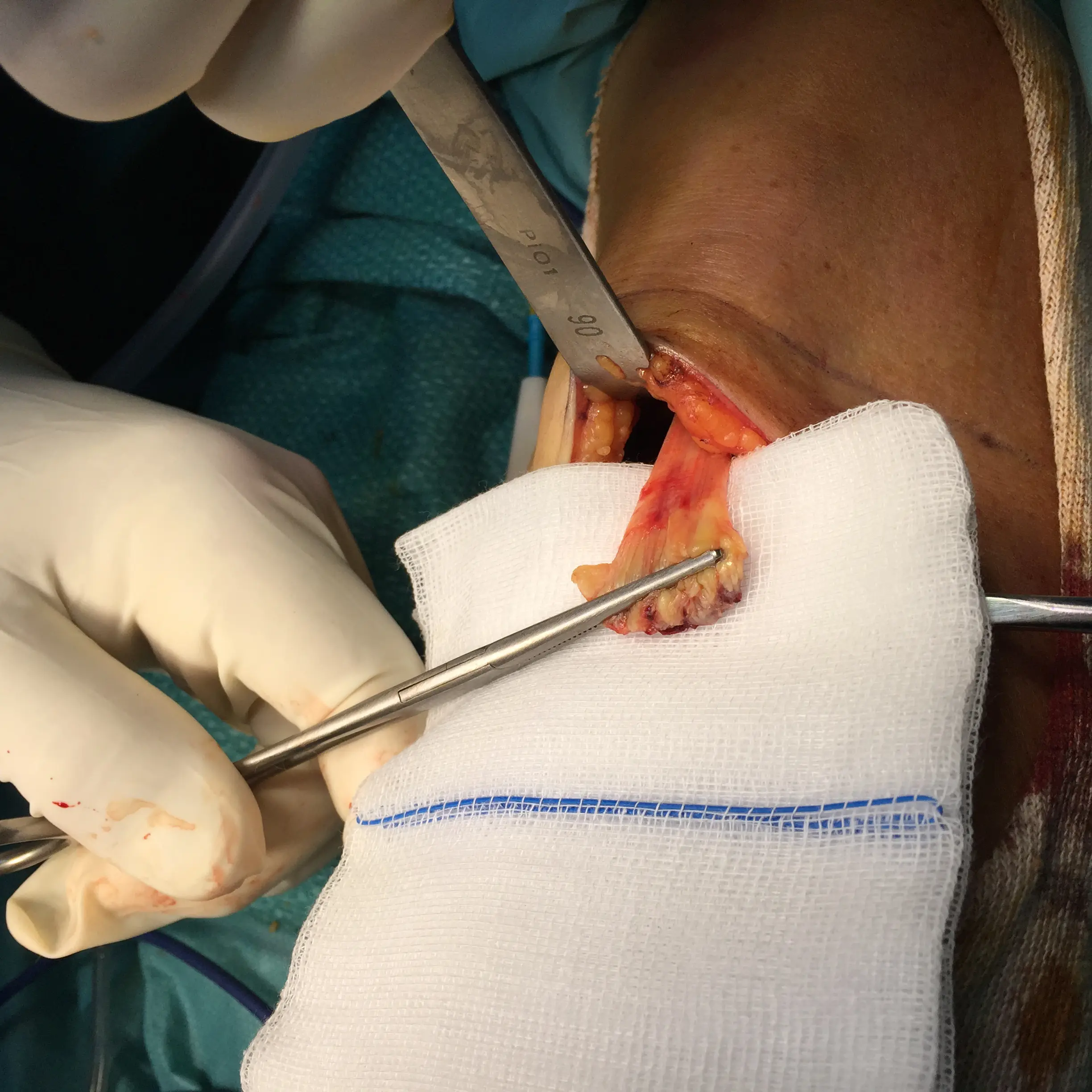
Treatment depends on the extent of the injury, ranging from taking anti-inflammatory drugs to local infiltrations with growth factors (PRP) to the need for surgery in the case of complete ruptures of the tendon.

Golfer's Elbow (Epitrochleitis)
LEARN MORE
DIAGNOSIS
This pathology can often be confused with the more common, “golfer’s elbow” or cervical radiculopathy. Therefore, it is always recommended to consult a specialist who, through careful physical examination and the aid of instrumental investigations, will be able to formulate a correct diagnosis. The diagnostic aspect is of particular importance as this clinical condition tends to become chronic especially in habitual players.
SYMPTOMS
The typical symptoms are dominated by pain, mainly located in the inner part of the elbow and exacerbated in some movements, sometimes even trivial, such as turning a door handle.
TREATMENT
The therapy consists of rest from sport, taking anti-inflammatory drugs, and using special adhesive bands or small braces to wear in during sports activity. In the case of more persistent forms, PRP infiltrations will be useful, as well as muscle stretching exercises and physical therapies. Surgery is rarely needed.
Osteonecrosis Of The Elbow
Osteonecrosis is the death of bone tissue due to a lack of or insufficient blood supply.
LEARN MORE
INJURY
Osteonecrosis is the death of bone tissue due to a lack of or insufficient blood supply.
Avascular necrosis, bone necrosis or bone infarction results in the appearance of tiny fractures in the affected bone tract.
The elbow is not frequently affected by this pathology, which may be the consequence of osteochondritis dissecans in adolescents (especially males) following a trauma or fracture of the capitulum humeri. It should not be confused with Panner’s Disease, which is an osteochondral disease of non-traumatic origin, typical of the first decade of life; this form is not evolutionary, does not generate intra-articular floaters, and undergoes spontaneous resolution with the decrease in sports loads.
CAUSES
The etiopathogenesis of osteonecrosis has a microtraumatic origin, due to trauma repeated over time, capable of determining not only a direct lesion of the osteochondral structures, but above all, the terminal vascular structures of the sub-chondral bone, determining their necrosis: it creates an intra-articular area unable to maintain structural stability and intrinsic functional characteristics.
DIAGNOSIS
It is necessary to resort to specific instrumental examinations: starting with an X-ray examination, which is useful in detecting bone changes that occur in the advanced stages of osteonecrosis; in the early stages, in fact, the problem is rarely identified. It is therefore better to use nuclear magnetic resonance imaging (MRI) which clearly shows the bone changes but is not always comprehensive at the beginning of the disorder.
A big advantage of MRI is that it is completely harmless to the patient.
SYMPTOMS
Elbow pain during flexion, extension, and pronosupination with functional limitation even in common activities.
TREATMENT
Treatment initially consists of rest, drug therapy, and physical therapies such as magneto-therapy.
Growth factors are used in the early stages of the disease.
However, it is a difficult pathology since healing is not always achieved.
In consideration of the early age in which the disease often manifests itself and, consequently, of the high functional demands, the importance of “biological” therapeutic options emerges which allow to spare the joint, avoiding bone collapse.
The research perspectives on which we have been focusing on for some years are aimed at the study of the pathophysiology of the disease and the use of autologous growth factors and mesenchymal cells that are proving to be fundamental in promoting the processes of reparative osteogenesis.
Patient Stories


Tendinitis of the biceps brachii