Spine
Learn about the most common spinal conditions that we can help you treat
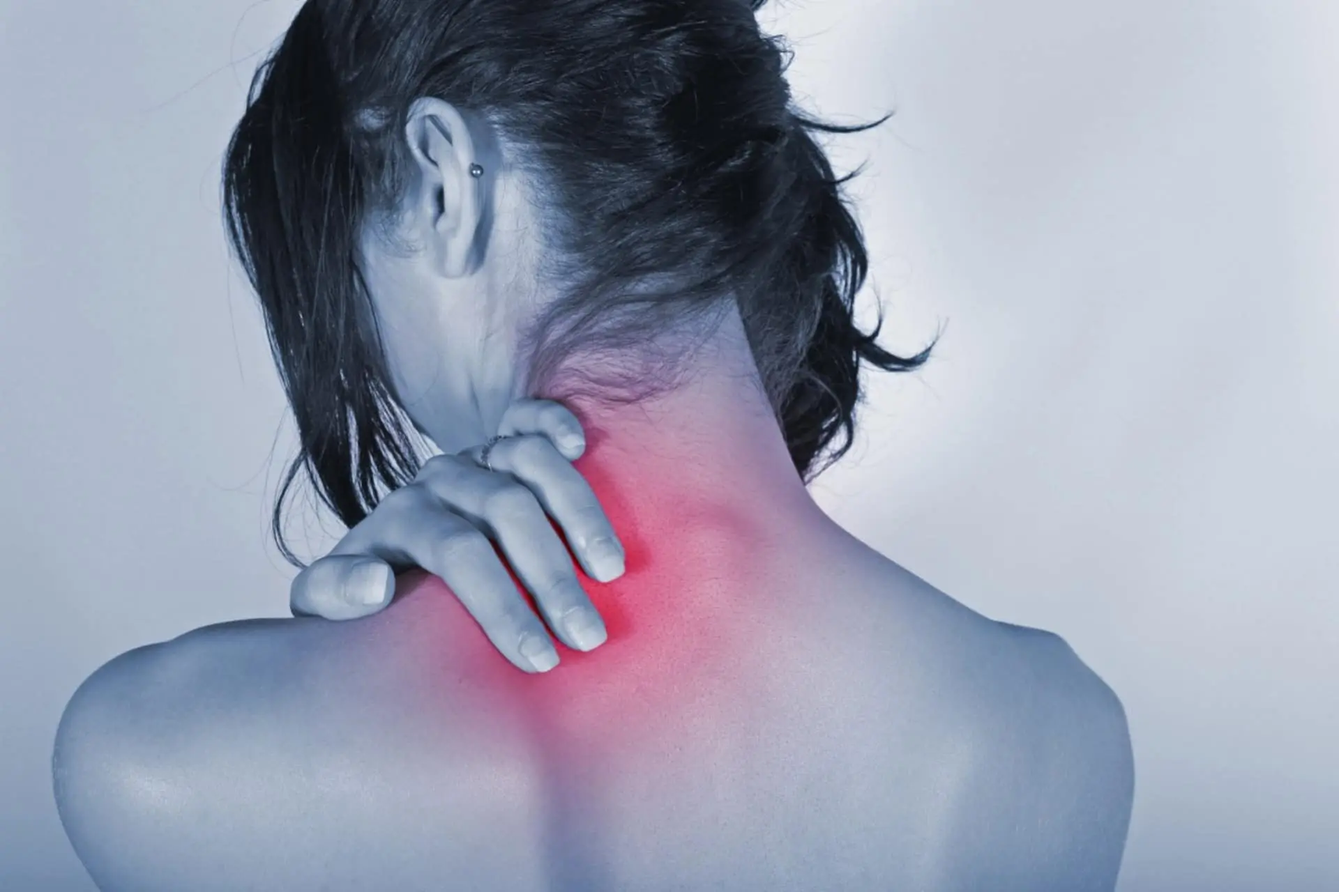
Cervical Spondylosis
Cervical spondylosis is a common degenerative disease of the cervical spine, usually expected beyond a certain age (60-70 years) due to degeneration of the cervical intervertebral discs.
LEARN MORE
INJURY
Cervical spondylosis generally appears in mature age and is due to the wear of the vertebral discs. In younger people, it is usually a consequence of trauma, diseases, or genetic factors.
Over the years, the discs can wear out and form bone spurs (osteophytes) on the vertebrae.
These protrusions can irritate the nerve endings of the spine and inflame the surrounding area, causing pain in the back and cervical area.
Correct posture, constant physical activity, and correct use of the back can significantly reduce the development of osteoarthritis.
CAUSES
The cervical spine may show a hypertrophic deformation of the vertebrae, with narrowing of the spinal and neural canals with possible compression of the nerve roots. This can cause pain that can radiate to the upper limbs, and limit mobility of the head with local pain.
Massive bone beaks (osteophytes) can form which can cause a decrease in the diameter of the vertebral foramina and a compression syndrome with radiculitis and marrow pain.
Cervical spondylosis usually happens at an older age and is due to wear and tear of the intervertebral discs. In the case of younger individuals, it is usually secondary to trauma, diseases, or genetic factors.
SYMPTOMS
It usually manifests as pain on movement, stiff neck, dizziness, headache, nausea, and stiffness. In the case of nerve root impingement, the pain may radiate to the upper limbs up to the hands also with tingling and poor or altered sensation. The following symptoms may also be present: sensation of dizziness in changes of position (lying down to upright), feeling of instability standing with eyes closed, and balance disturbances.
TREATMENT
- Cervical spondylosis therapy ranges from the use of painkillers such as NSAIDs, paracetamol, cortisone, muscle relaxants, and neuro protectors.
- Another effective treatment is PRP which, being rich in growth factors and anti-inflammatory substances, can help mitigate the problem.
- Much importance is attached to FKT and the use of local physical therapies such as laser and TECAR therapy, application of a collar, osteopathic treatments, postural gymnastics.
- If severe neurological deficits occur, surgery may be necessary with surgical decompression, laminectomy, stabilization, or application of intervertebral or joint prostheses.
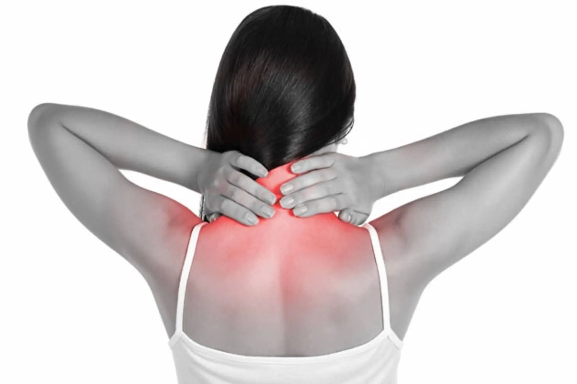
Neck Pain
One of the most common problems in the Western world. Most often the cause is cervical inflammation which affects the upper vertebrae of the spine, those that support the neck and head.
LEARN MORE
INJURY
Pain causes stiffening of the neck and a strong decrease in movement.
It often tends to radiate to the shoulders and arms and is accompanied by other symptoms such as headache, dizziness, nausea and sensory disturbances (sight and hearing).
CAUSES
Common causes are incorrect postures, poor exercise, stress, osteoarthritis, or acute trauma (injuries during sports, whiplash)
In order not to create problems, the neck must be elastic enough to move without causing pain, in 6 different directions:
Flexion and extension (to move the head forward and backward);
Rotation (head moves left and right);
Lateral tilts (the head is brought with the ear toward the corresponding shoulder)
SYMPTOMS
The neck pain perceived by patients is often variable. It is a constant pain that can be localized in different areas of the cervical spine.
Localization often helps to understand the origin of the problem, whether or not it involves the nerves and if perhaps an inflammatory reaction is underway.
In most cases, the pain does not last longer than a few days and rarely exceeds beyond a week, especially when they are of traumatic origin. They resolve spontaneously, or with the help of over-the-counter drugs.
If not treated properly, however, the symptoms tend to recur with relative ease.
PREVENTION
Diagnosis: The diagnosis of sciatic nerve pain is mainly linked to the anamnestic collection (history of the disease, onset and characteristics of the symptoms) and to accurate and focused clinical observation.
Effective instrumental tests for confirming the diagnosis of lumbosciatica are: x-rays of the lumbosacral spine, MRI, or CT scan.
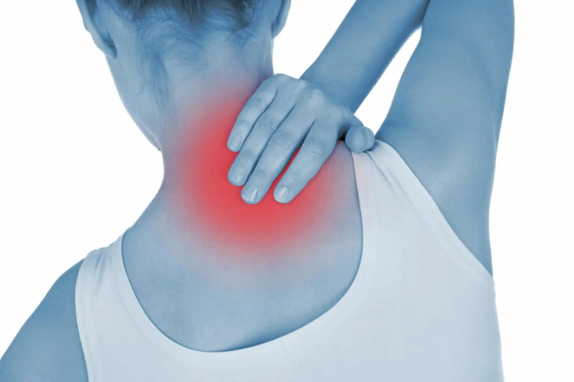
Whiplash
Whiplash is a traumatic event that occurs mostly as a result of traffic accidents, typically anteroposterior rear-end collisions, in which the patient’s body is thrust forward by the impact and the neck is stretched as a result of skull inertia.
LEARN MORE
CAUSES
It is due to a strong stress on the vertebral, ligament and muscular structures of the neck caused by the whiplash and can give rise to pathological consequences such as contractures of the paravertebral muscles, reduction of the physiological lordosis of the cervical tract, muscle tear, crushing or fracture of the cervical vertebrae.
SYMPTOMS
The result of all these harmful effects translates into the classic whiplash symptomatology, generally characterized by:
- neck pain;
- ringing in the ears;
- stiffness of the back and neck muscles;
- headache;
- movement restrictions.
TREATMENT
- Rest is certainly the most effective means of healing, limiting the movement of the neck and head as much as possible. Often, in fact, one of the first solutions adopted is the application of the collar, which supports and protects the cervical tract.
- Thanks to the immobility and the combined use of adequate drugs (FANs, muscle relaxants or sedatives), cervical inflammation can recede in reasonable times.
- Physical therapies can also help speed up the healing process.
RECOVERY
.
.
.
Disc Problems
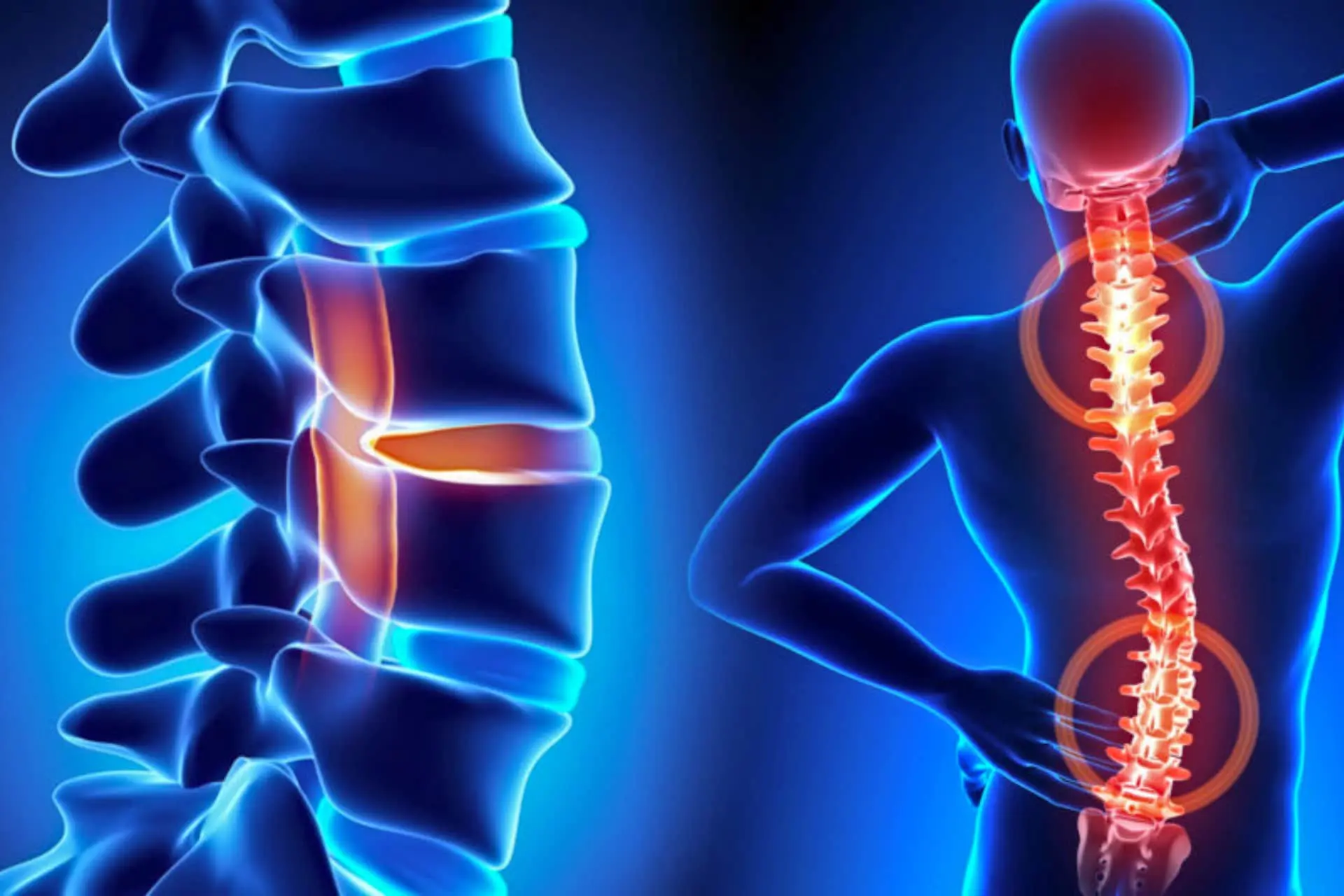
Disc problems refer to conditions such as herniations, protrusions, or degeneration of the disc.
LEARN MORE
The most common back problems originate in the discs: it can tear, form a hernia, or wear out (degenerate).
In all these cases, it becomes difficult for the discs to perform its task to act as shock absorbers for the vertebrae. The area around the vertebra can thus become inflamed and the nerve endings can become irritated.
The muscles then contracts (spasms) to protect the spine, increasing the sensation of pain.
Annular Tear
The annulus fibrosus is the very resistant outer part of the discs. A sudden movement can cause a micro-laceration inside a disc and the surrounding ligaments could undergo an elongation stress.
Since the annulus fibrosus and ligaments contain nerve endings, pain may be elicited.
Disc Protrusion (Herniated Nucleus Pulposus)
As the disc weakens, the jelly-like inner part calle the nucleus pulposus can be compressed against the annulus fibrosus, which can force the disc out of its seat (protrusion). This irritates the spinal nerve roots causing back and leg pain.
In more serious cases, surgery may be required to resolve the problem.
Slipped disc
If the fibrous ring is torn, the nucleus that was already pushing against its outermost wall comes out and compresses the nerve ending of the spinal column.
If the resulting pain spreads to the back of the thigh and leg, it is called sciatica. When the compression on the nerve root is very strong or lasts over time, they can be damaged and cause numbness and weakness in the leg muscle, sometimes accompanied by severe pain and disability.
In more serious cases, surgery may be required. During the operation, the doctor may remove the part of the disc that irritates the nerve root (discectomy) after opening a window in the back of the vertebra (laminectomy or laminotomy).
In other cases, the surgeon might join adjacent vertebrae together (fusion) to make the spine more stable.
Coccyx Fracture
Who hasn’t slipped and landed on their behind at least once in their life?
It usually ends with a little pain and a bruise but unfortunately sometimes our “tail” breaks and we have a coccyx fracture, often difficult to detect with conventional radiology since our last vertebrae have a morphology that varies from individual to individual.
LEARN MORE
INJURY
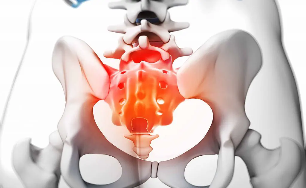
In these cases, the clinical experience helps more, and an accurate clinical examination and a targeted CT scan is requested if a fracture or dislocation is suspected. Let’s try to understand something more about this area: the coccyx represents the terminal tract of the vertebral column, located just above the intergluteal cleft. It articulates with the sacrum and consists of four fused vertebrae together and excluding the first. The terminal apex is slightly curved forward so that the apex of the bone is oriented toward the front of the body. The dorsal surface of the coccyx serves as an attachment site for the gluteus maximus, the pubo-coccygeal muscle and the ano-coccygeal ligament, while toward the apex the anus sphincter inserts.
CAUSES
Coccyx injuries are generally associated with a direct fall on the buttocks, and the patient describes intense pain that appears upon sitting or during bowel movement. In most cases, only a contusion will arise however if the trauma is severe, it can cause a fracture or dislocation (i.e. a displacement from its natural location) of the coccyx. If this happens, its component bones can become hypermobile, triggering pathological instability that manifests as chronic inflammation and pain. The diagnosis of a coccyx fracture is made with a rectal examination, evoking pain by moving the coccyx. Radiographs are not essential for diagnosing coccygeal fractures.
SYMPTOMS OF COCCYGODYNIA
The main symptom of the disorder is pain localized at the base of the spine, where the coccyx is located. The pain can be so intense that it makes it difficult for you to perform normal daily activities, such as driving, bending over, or sitting down. However, its intensity varies from person to person, and can worsen over time. Pain in the coccyx can be particularly intense when the patient rises from a sitting position or when the region is directly compressed.
TREATMENT
Treatment of coccygodynia varies depending on the underlying cause and extent of pain; usually, it responds well to conventional therapy with nonsteroidal anti-inflammatory drugs (NSAIDs) to relieve pain, combined with muscle relaxants to relieve excessive muscle contraction. The first approach involves the administration of analgesics/anti-inflammatories, such as ibuprofen, naproxen, paracetamol, or tramadol. Sometimes, if the pain is particularly intense, local infiltrations can be performed and PRP can help in the consolidation of a fracture. Symptoms usually resolve within a few weeks but for those who practice activities where they have to stay in a seated position subjected to possible trauma, the times are longer.
Some osteopathic spinal manipulation techniques can provide pain relief.
In a small number of cases, surgical removal of the coccyx (coccygectomy) may be recommended but only when all other conservative treatments have failed. Surgery is usually reserved for patients with advanced coccygeal instability (e.g., subluxation or hypermobility). During a coccygectomy, the surgeon makes a small incision to completely or partially remove the tailbone. Muscles, tendons and ligaments attached to the excised bone segment are reconnected to other parts of the pelvis to preserve their function. Surgical therapy is effective, but local postoperative infection may occur in about 30% of patients due precisely to the proximity to the anorectal area.
Low Back Pain
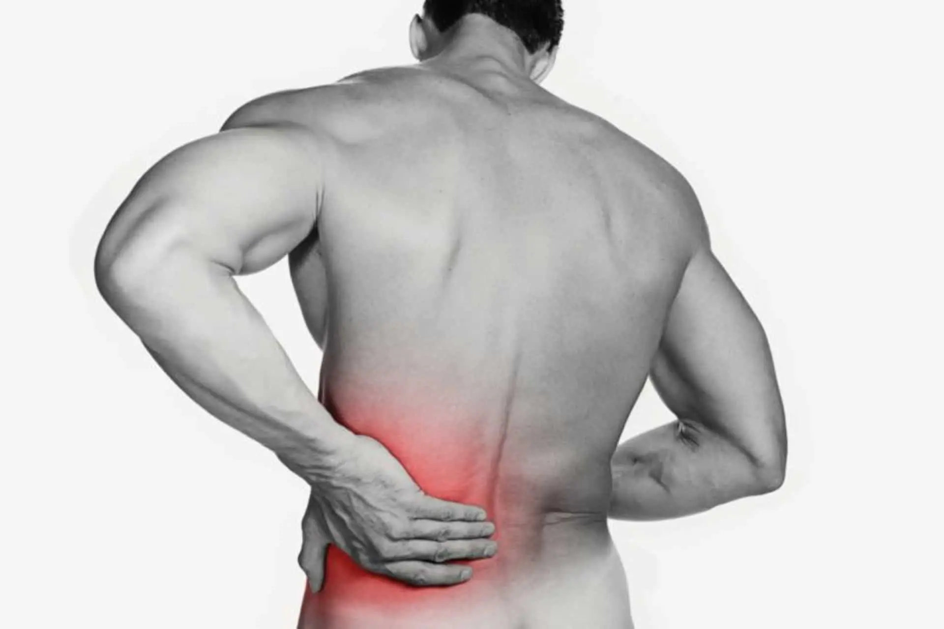
Low back pain is one of the most common ailments, as everyone suffers from it at a certain stage of active adult life.
It often appears suddenly, without an apparent cause, and can interfere with simple daily activity, movement and sleep, and then disappears just as suddenly.
LEARN MORE
CAUSES
Acute low back pain can have various causes.
In most cases, it is due to an injury of the muscles or ligaments belonging to the back; more rarely, it is the result of a vertebral fracture, vertebral tumor, herniated disc, vertebral stenosis, sciatica, state of pregnancy, etc.
Very often, the presence of acute low back pain is associated with other symptoms, such as stiffness, difficulty moving and tingling.
SYMPTOMS
The pain related to acute low back pain may be piercing, burning or dull; moreover, it may be continuous or alternate between phases of great intensity and phases of apparent remission.
Very often, the pain that characterizes acute low back pain is associated with other symptoms, including:
- Stiffness in the lumbar region;
- Tingling or burning in the lower back;
- Difficulty in movement, albeit minimal, due to exacerbation of pain.
- Additionally, depending on the underlying cause, the patient may also experience:
- Pain, tingling and / or weakness in a thigh, leg and / or foot;
- Lameness;
- Loss of control of the anal or bladder sphincter;
- Fever;
- Weight loss for no reason.
DIAGNOSIS
For the diagnosis of acute low back pain and the recognition of its underlying cause, objective examination and history are sufficient.
If, however, the objective examination and medical history are not sufficient for a definitive diagnosis, instrumental tests of the spine, such as X-rays, MRI or, in some instances, even CT scans, must be used.
TREATMENT
Since acute low back pain is most often due to a muscle or ligament injury, treatment includes:
- Rest from any activity that may accentuate the pain;
- Ice application;
- Taking anti-inflammatory drugs;
- Correct posture.
Sciatica
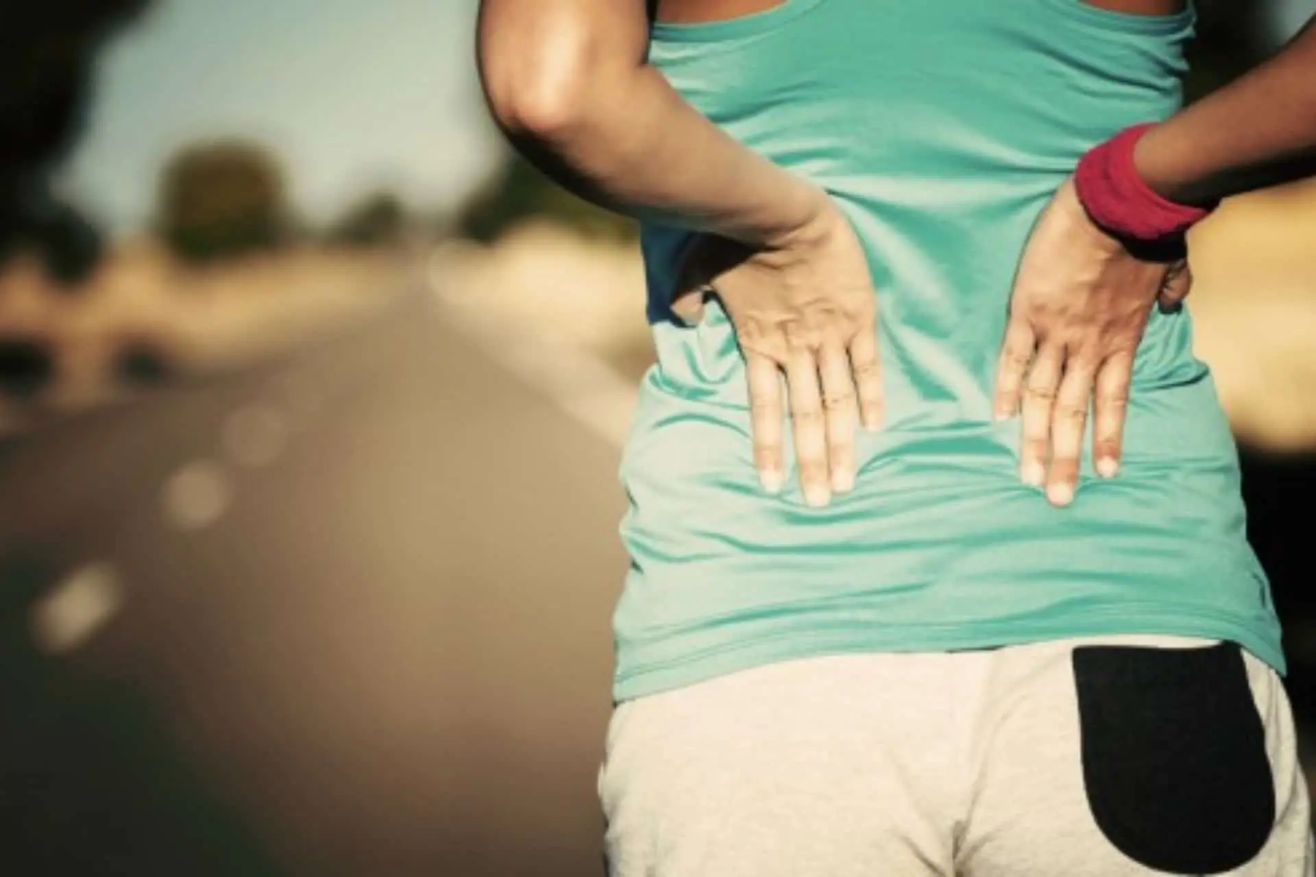
Sciatica is characterized by pain in the lumbar or lumbosacral spine, radiating to the lower limb where the sciatic nerve is located.
LEARN MORE
INJURY
The pain can be localized both posteriorly (gluteal region, posterior aspect of the thigh and leg) towards the sole of the foot, or radiate to the lateral part of the thigh and leg towards the back of the foot and the big toe.
Sciatica pain caused by nerve compression (radiculopathy) is often described as sharp, stabbing, relatively well defined, and similar to an electric shock. There may be concomitant alterations in sensitivity such as tingling, numbness, reduction in the strength of some muscle groups of the leg, foot, thigh and consequent difficulties in walking.
CAUSES
Causes of low back pain can be:
- heavy work
- weight lifting
- repetitive twisting of the trunk
- pathological changes in the curve of the spine
- any previous spinal surgery (hernia recurrence).
SYMPTOMS
Symptomatology may start suddenly with disabling character and occur following an abrupt movement or even without apparent cause. Symptoms may continue acutely for several days and then gradually subside.
DIAGNOSIS
The diagnosis of sciatica is mainly related to history collection (history of the disease, onset, and characteristics of symptoms) and accurate and focused clinical observation.
Effective instrumental tests for confirming the diagnosis of lumbosciatica are: x-rays of the lumbosacral spine, MRI, or CT scan.
TREATMENT
- Progressive cautious mobilization and pharmacological treatment (anti-inflammatory and muscle relaxants), are indicated. In resistant cases the use of cortisone is preferred.
- When the pain and related symptoms subside, it is necessary to start a rehabilitation process whose objectives are aimed at: identifying the cofactors involved in the genesis of sciatica, rebalancing the posture of the pelvis, lumbosacral tract and lumbar spine.
Scoliosis
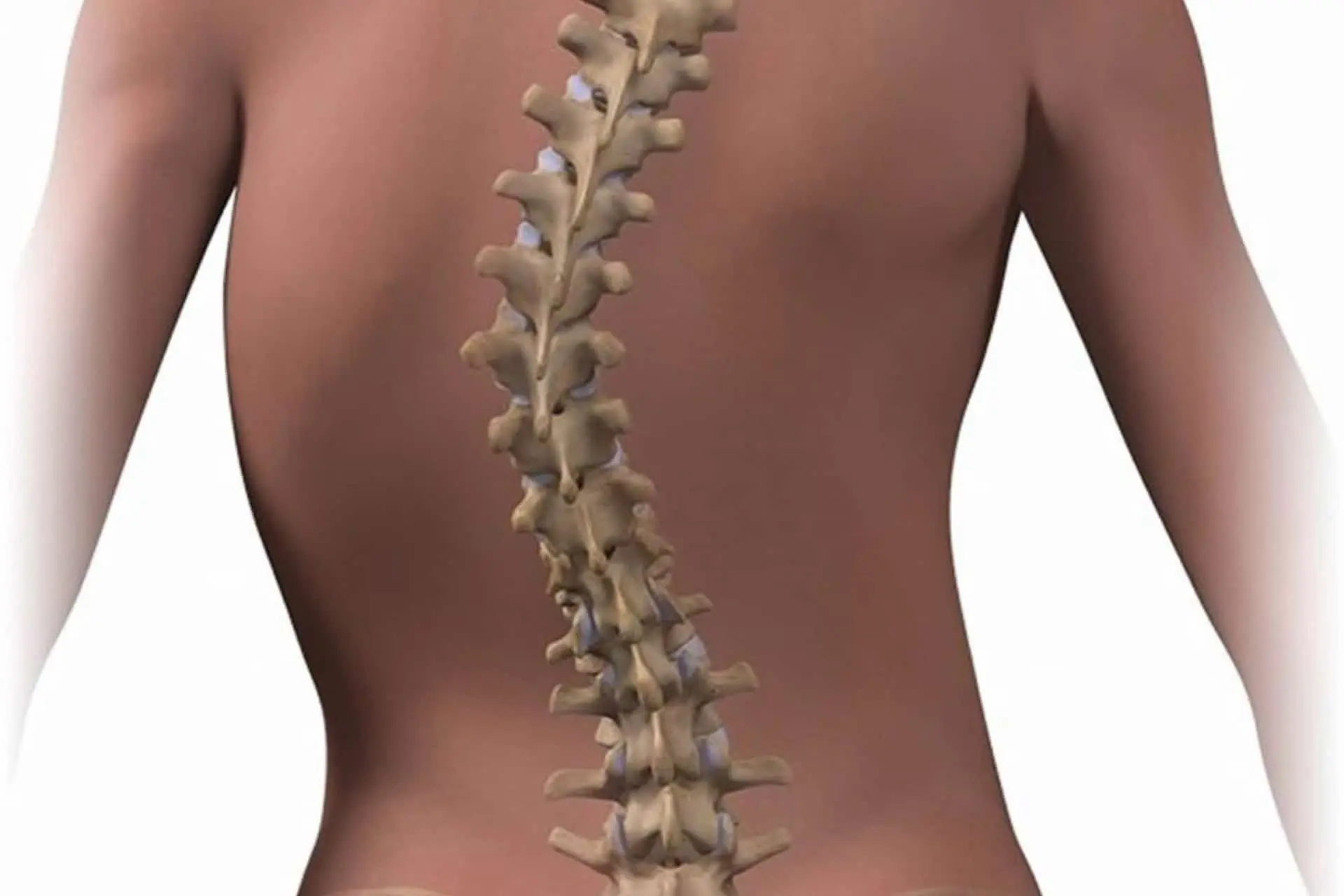
The term scoliosis refers to an abnormal curvature of the spine.
LEARN MORE
CAUSES
It can be congenital or develop in adolescence.
The mild forms cause only slight discomfort, but the more severe ones generate chronic pain and can affect internal organs.
Diagnosis is based on physical examination and radiographs.
Not all forms of scoliosis progress, but if they do, they need to be treated as soon as possible to prevent severe deformities. Braces or surgery are used to realign the spine.
The disorder is relatively common and occurs in 2-4% of children between 10 and 16 years. Males and females are equally affected. However, it is 10 times more likely to progress and require the use of braces in females.
Scoliosis can have hereditary causes, congenital defects or injuries or develop over the course of life, usually at the beginning of adolescence.
Usually the cause cannot be identified (so-called idiopathic scoliosis).
The spine flexes to the right if the defect is in the back and to the left if the defect is in the lumbar region.
The right shoulder is typically higher than the left and the pelvis may be misaligned. The chest can also be asymmetrical.
SYMPTOMS
Mild forms of scoliosis are usually asymptomatic. Sometimes, the child has pain or stiffness in his back if he is sitting or standing for a long time. Over time, the pain can worsen to intense.
Several factors contribute to the evolution of scoliosis.
The steeper the curve, the more likely the deformity is to get worse, particularly in the early stage of puberty when growth is accelerated.
Likewise, the more prominent the symptoms, the greater the likelihood that scoliosis will progress.
In the long run, the aggravation of the condition can cause permanent problems, such as obvious deformities or chronic pain.
Severe forms can also have an effect on internal organs, for example by deforming and damaging the lungs.
DIAGNOSIS
To diagnose it, the doctor asks the child to lean forward and observes the spine from behind, because from this position it is easier to detect any deviations.
The radiographs allow you to accurately detect the angle of curvature.
If the doctor suspects progressive scoliosis, he may ask to see the child several times a year. There are special techniques for measuring the curves of the spine.
PROGNOSIS
In most children with scoliosis, the deformity does not progress and remains contained. However, it is important that your doctor examines it regularly.
Symptomatic, progressive or severe scoliosis must be treated. The earlier treatment is started, the greater the chance of preventing a severe deformity.
TREATMENT
- Corset and physiotherapy
- Sometimes surgery
- Both the affliction itself and the treatment can harm self-esteem and body image. For this, psychological support or psychotherapy can be useful.
Ankylosing spondylitis
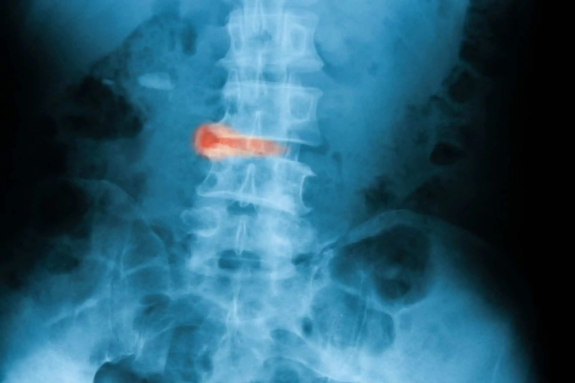
Ankylosing spondylitis is an inflammatory pathology of the spine, which involves the sacroiliac joint and can occasionally involve other organs such as the eye (anterior uveitis).
LEARN MORE
CAUSES
Unleashed is unknown; what is known is that it has an autoimmune genetic substrate, linked specifically to human leukocyte antigens, more specifically to HLA-B27.
This syndrome is 3 times more frequent in males than in females, with an age of onset of less than 40 years.
SYMPTOMS
One of the earliest manifestations of ankylosing spondylitis is sacroiliitis, which manifests as morning stiffness localized to the lumbosacral spine.
This stiffness never lasts longer than 30 minutes, a characteristic that places it in the differential diagnosis of rheumatoid arthritis. Pain-related sleep disturbance is common.
Other frequent manifestations are localized stiffness and pain in the major joints (hip, shoulder), present in 30/40% of patients with ankylosing spondylitis.
DIAGNOSIS
Diagnosis is usually radiographic, supported by the patient’s medical history and laboratory tests.
TREATMENT
- The treatment is multimodal.
- Maintaining an adequate level of physical activity is of fundamental importance.
- Physical therapies (FKT) and the use of non-steroidal anti-inflammatory drugs, possibly associated with muscle relaxants, may be envisaged.
- A visit to a specialist is good practice, due to the difficult differential diagnosis and the correct classification, so as to be able to start the correct therapy.
Spondylolisthesis
Spondylolisthesis is a degenerative pathology of the lumbar spine, which manifests itself with pain and functional disability. It is more common in women, especially after 40 years of age.
LEARN MORE
INJURY
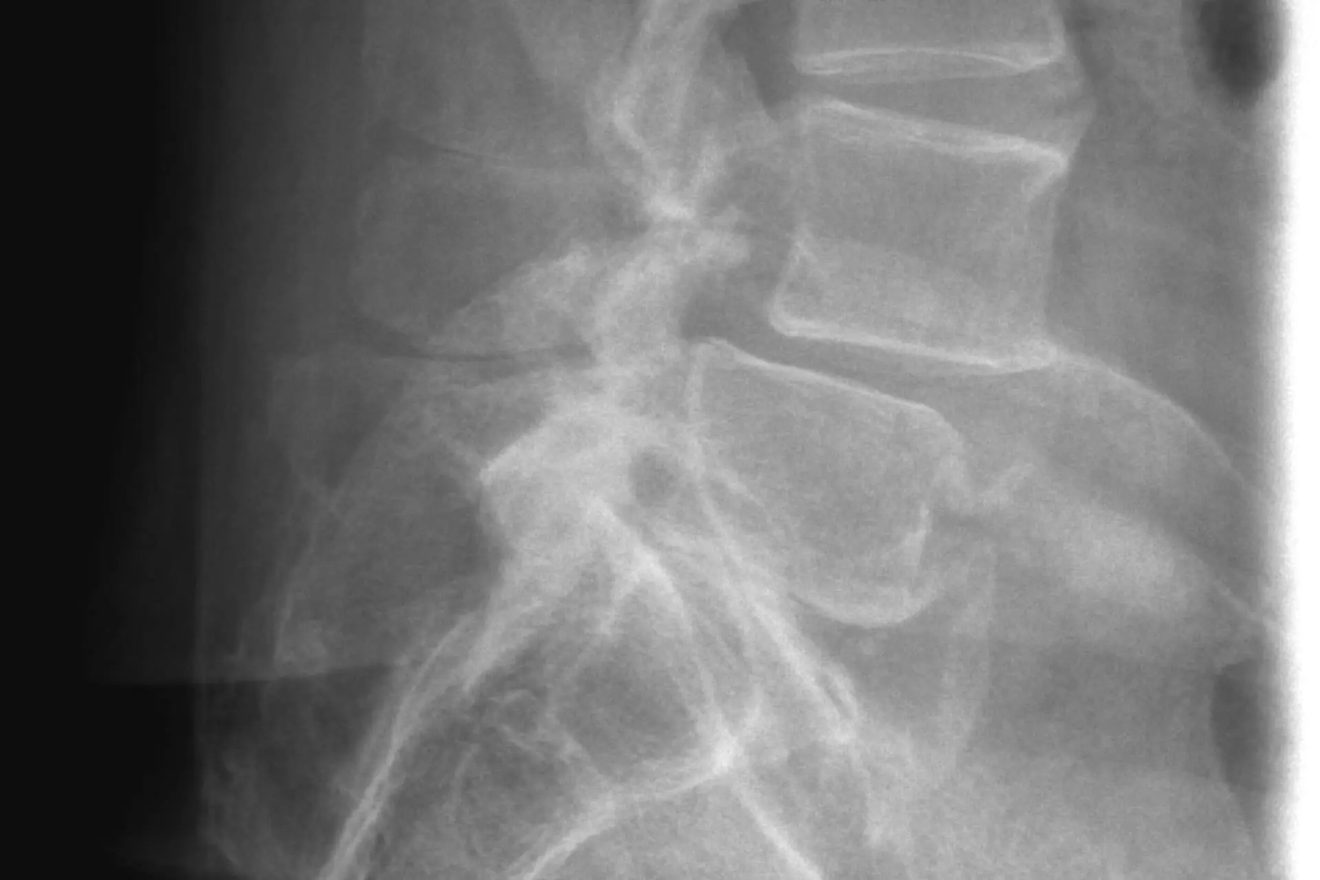
This pathology is characterized by the slippage of a vertebral body against the underlying one, due to the degeneration of the articular facets and the vertebral disc.
This slippage can result in a constriction of the spinal cord which leads to the appearance of pain.
Often, the pain is radiated to the lower limbs, resulting in a lameness during walking and is accentuated in the transition from standing to sitting and vice versa.
CAUSES
There are several causes that can give rise to this spinal disorder:
- presence of previous spondylolysis (rupture between the body and the posterior arch of the vertebra);
- deterioration of the vertebra and its disc;
- trauma to the spine;
- infections (Pott’s disease, also called tuberculous spondylitis or vertebral tuberculosis).
SYMPTOMS
Symptomatology is proportional to the extent of vertebral displacement and is manifested by the typical low back pain aggravated by physical exertion and various complaints in the lumbosacral region.
The pain of spondylolisthesis can be made worse by standing for long periods of time.
In severe cases it can become chronic and be accompanied by pain typical of sciatica.
DIAGNOSIS
A visit to a specialist is always recommended, in order to set up an adequate diagnostic-therapeutic procedure. The clinical diagnosis must be supported by imaging (X-ray, MRI, CT) of the lumbar spine.
TREATMENT
- The treatment is multimodal and includes physical therapies, analgesics, infiltrations.
- In addition, absolute bed rest is recommended until the painful symptoms are resolved and the intake of analgesic and anti-inflammatory drugs has ceased
- The practice of postural gymnastics is also important, but not during periods of acute pain.
PREVENTION
The prevention of spondylolisthesis is in turn based on the prevention of conditions that can lead to the development of the disease.
It is therefore advisable to avoid overloading the spine excessively, in order to minimize the risk of deterioration or breakage of the vertebrae and, for the same reason, to avoid trauma to this delicate part of the body as much as possible.
Tietze Syndrome
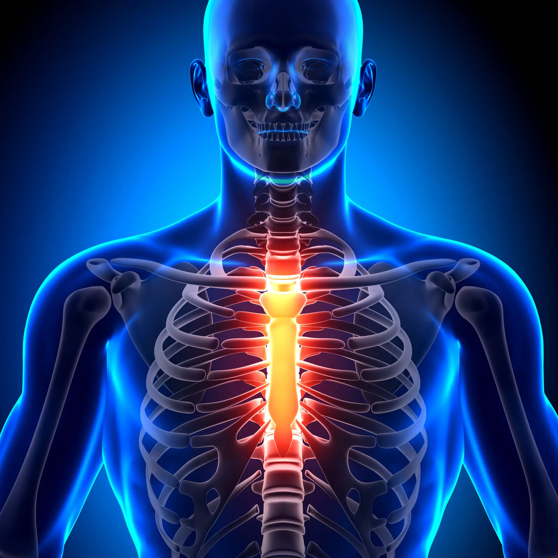
La sindrome di Tietze è definita come un’infiammazione benigna, dolorosa, self-limiting, non-suppurative and localized inflammation of the upper costal cartilages. It has unknown etiology. It affects the costochondral, costosternal, or sternoclavicular joints. The manubrium-sternum and xiphosternal joints are less frequently affected.
LEARN MORE
Although they were once considered separate conditions it is now recognized as a severe form of costochondritis. Tietze’s syndrome differs from the less severe forms of osteochondritis by the swelling of the costal cartilages.
CAUSES
The underlying cause of the onset of Tietze’s syndrome is currently unknown, but various hypotheses have been formulated. The most accredited theory assumes that small traumas (so-called micro traumas) to the chest wall, over time, can cause damage and consequent inflammation to the ribs.
These micro traumas, not severe enough to be visually highlighted, can result from a sudden event, such as a car accident or surgery, or develop following excessive exertion such as repeated cough and vomitingover time.
SYMPTOMS
Clinical manifestations include sudden or gradual onset of pain of varying intensity in the anterosuperior wall of the chest, in association with a swelling of the costal cartilages, fusiform in appearance and soft on palpation. Despite the possibility that the pain may radiate to the shoulder, arm, and neck, its distribution is usually within the segment innervated by the afferent fibers carrying the pain impulse.
Pain is often aggravated by chest wall movement, sneezing, coughing, deep breathing, bending, straining, and prone position. Some patients report an inability to find a comfortable position in bed and have pain when turning over in bed; weather changes, anxiety, worry, and fatigue can exacerbate symptoms. The pain is usually unilateral, without a preferential hemithorax. No association with sternotomy is reported.
TREATMENT
To date, there are no guidelines to refer to for identifying a unique therapy for Tietze syndrome; for pain relief, the so-called conservative methods are the first line of treatment to be used, which include rest for a short period of time with abstention from physical activity.
Analgesic drugs such as acetaminophen or nonsteroidal anti-inflammatory drugs(NSAIDs) may be prescribed in association. The use of ice packs placed on the affected area can also be useful in relieving pain.
In the event that the aforementioned drugs do not bring the expected benefits, you can opt for other solutions, one of the most effective has been shown to be the injection of lidocaine (used as a local anesthetic) in association with corticosteroids. The latter, despite being among the most effective drugs when taken orally, can present, especially if used for long periods, various side effects, especially for people already affected by other chronic diseases. For this reason, therefore, there is a tendency to prefer its use by local injection.
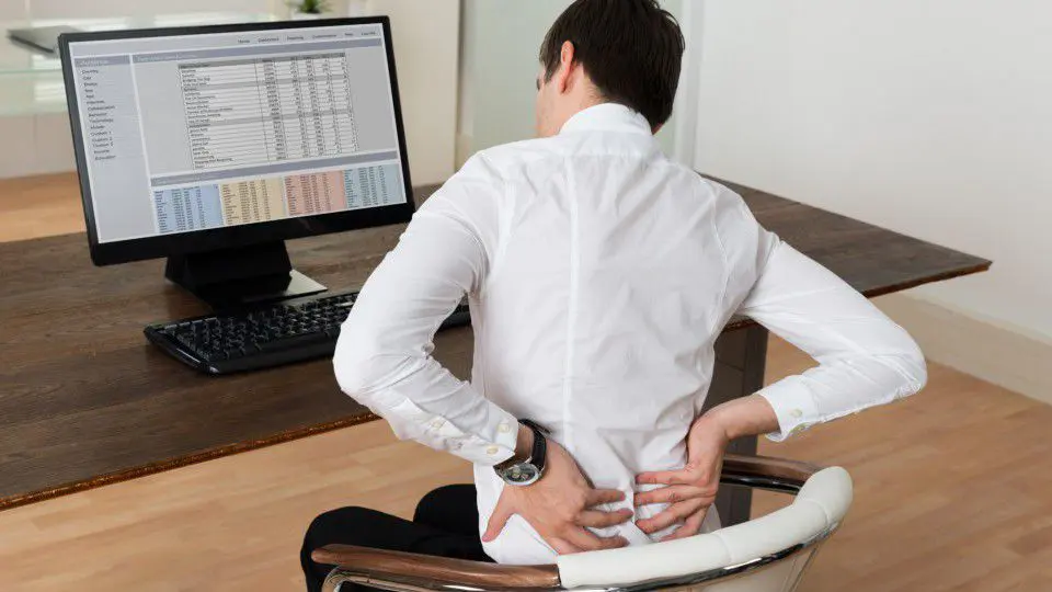
Backache
Back pain is discomfort or stiffness in the spine, often caused by muscle strains, disc problems, poor posture, or pre-existing medical conditions.
LEARN MORE
BACKACHE
Back pain almost always depends on a functioning difficulty of one of its tiny components (muscles, ligaments, disc). The back is like a high-precision watch in which even a very small grain of sand (a small injury) can cause considerable difficulty (severe pain). However, there is also a big difference: the back is enormously more solid than a watch. In fact, the spine is one of the strongest structures in our body, which has to perform many important tasks: stability: supports the trunk, upper limbs and head; mobility: allows all movement of the trunk and head; and containment: protects the spinal cord.
These functions contradict each other (stability-standing still is the opposite of mobility-moving). So this structure is strong but also extremely complex, with a very sophisticated control system. Its functioning depends on the one and only “on-board computer”: the brain, which manages the proper balance of the forces being unloaded on it. Loss of this balance implies the appearance of pain, which signals that the back is “out of control,” “out of shape.”
INJURY
Since we evolved from a position on all fours, the lower part of the spine has had to undergo a series of adaptations for which it was not initially designed: this probably led to the appearance of back pain, a pain typical of the human race. Having now forced the lower back to adapt to a sitting position it has further increased the problems. This is why everyone suffers from back pain sooner or later: it is our lifestyle as men, but also the lifestyle of each of us individually plays a fundamental role.
CAUSES
The theoretical causes of back pain are very numerous. Even more are the medical terms that indicate these causes. There are different words that are often different definitions of the same pathology: this is why going to many doctors often means being assigned as many diagnoses.
Serious causes of back pain are very rare (less than 1 in 300 cases) and a medical examination is enough to identify them. It is important, therefore, not to panic: everyone, sooner or later, suffers from back pain.
TREATMENT
Nine out of ten patients recover within a month of the onset of pain, regardless of whether or not they have had treatment: so it is not always necessary to rush to the specialist at the first signs of back pain. However, even if it heals on its own the pain should not be ignored: even if linked to almost always a trivial cause, relapse is, in fact, often frequent. This is precisely what makes prevention essential. In any case, relapse is not a sign of something serious, but only of the recurrence of a problem we are predisposed to. Treatment with drugs, physical or manual therapies is not always decisive, although it is often a valid help. Even if the pain does not go away, it does not mean that the situation is serious: in fact, there is no correlation between the amount of pain and the amount of damage. However, the therapies are only needed in case of acute pain, but have no preventive effect.
PREVENTION
- Exercise regularly and avoid sitting for too long.
- Do not smoke.
- Maintain good posture.
- When lifting weights, keep your back straight and bend your legs.
- Use a mattress that suits you, neither too soft nor too hard.
Patient Stories