Hip
Learn about the most common hip conditions that we can help you treat
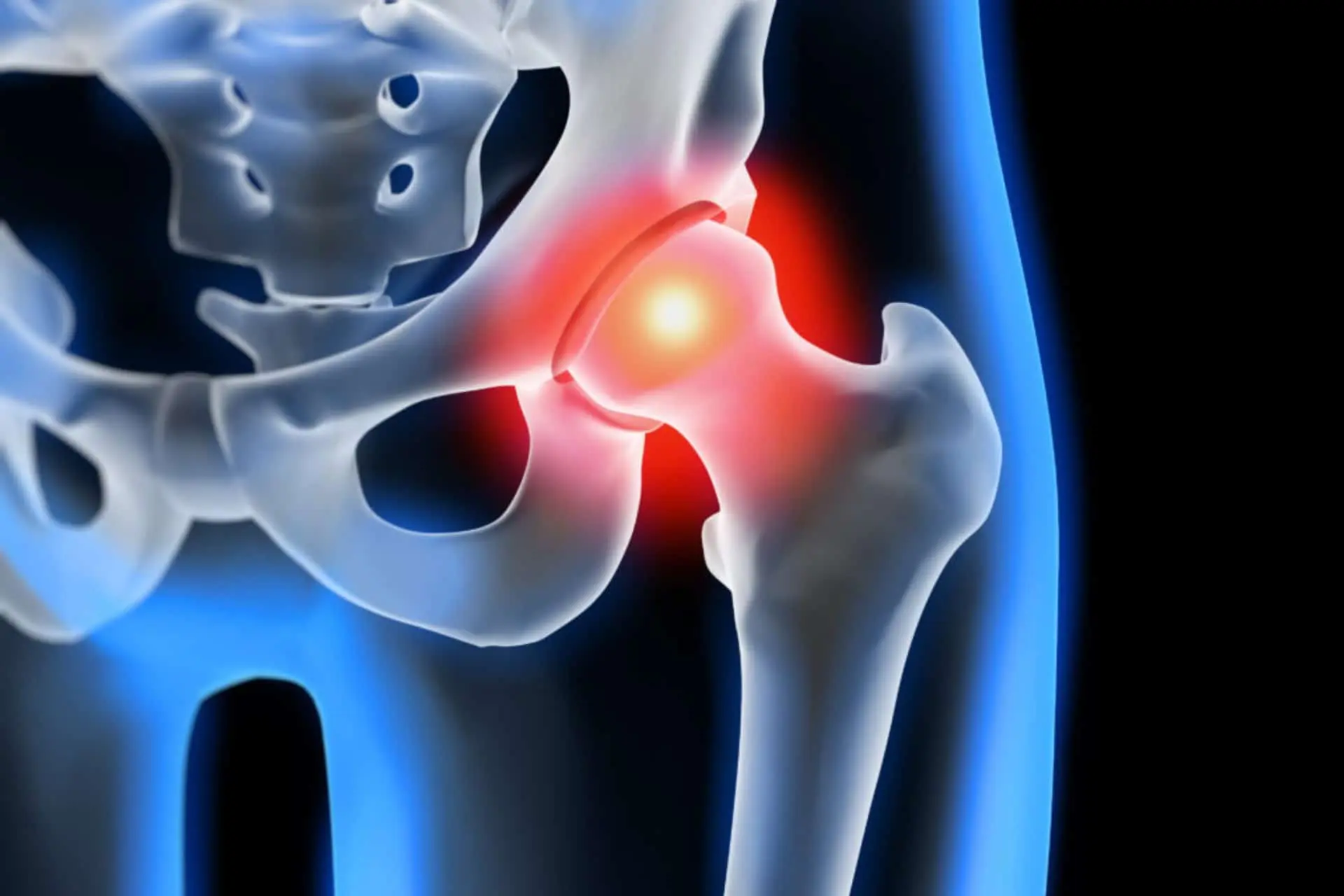
Hip Pain - Coxalgia
Coxalgia simply means hip pain, especially of the femoral – acetabular joint.
LEARN MORE
INJURY
There are several conditions, more or less serious, that can lead to coxalgia:
- Hip osteoporosis
- Avascular necrosis of the femoral head
- Other more rare, benign or malignant conditions, such as trauma, osteomyelitis etc.
CAUSES
Coxalgia with functional limitation can be a symptom of a numerous and varied set of idiopathic or degenerative, infectious, traumatic, neoplastic pathologies.
The differential diagnosis between the various diseases that cause coxalgia is extremely complex.
The most economical and common diagnostic procedure, namely that of the clinical examination followed by an X-ray examination, is not always definitive in this pathology, in which both clinical and radiographic signs are not pathognomonic.
Some elements collected in the anamnesis can facilitate the formulation of a differential diagnosis.
SYMPTOMS
Osteoporosis of the hip begins with dull pain in the groin region, in the buttocks, or in the anterior region of the thigh, with an acute beginning, accompanied by lameness and antalgic gait.
Patients are often able to report the exact date on which symptoms appeared. The pain is exacerbated by load and subsides at rest. Only rarely is it present at night.
In avascular necrosis of the femoral head, the onset of symptoms is more insidious, the pain exacerbated by load and does not subside at rest.
The antalgic gait appears later and there is a constant proportion between the level of pain and joint function.
DIAGNOSIS
- Radiography
- Bone scan
- Magnetic resonance imaging
TREATMENT
Treatment depends on the diagnosed pathology and may involve simple administration of anti-inflammatory drugs, PRP infiltration, up to surgery. We therefore recommend that you contact a specialist.
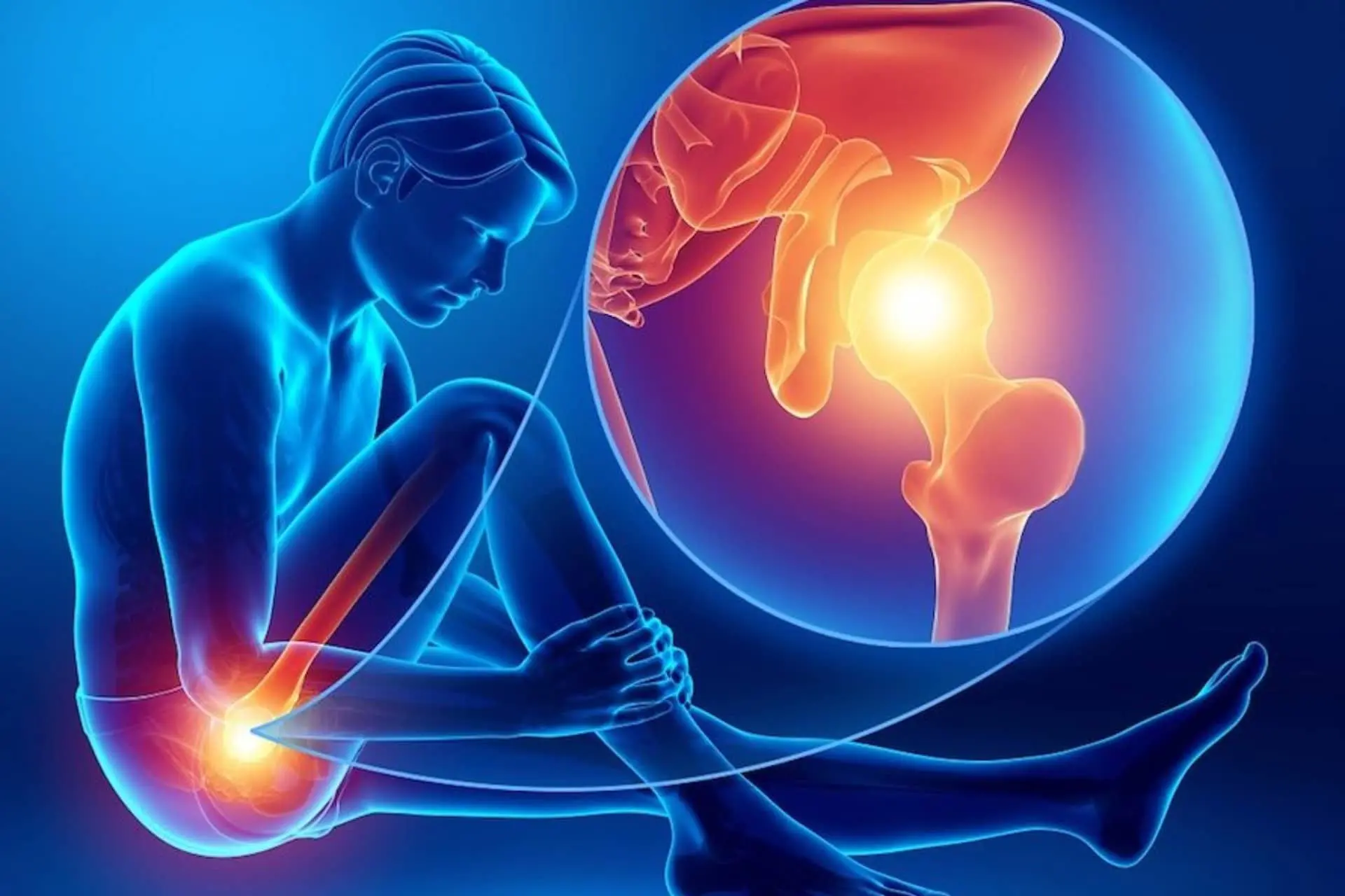
Femoroacetabular Impingement
Femoroacetabular impingement is a condition in which abnormal bone growth in the hip joint causes friction.
LEARN MORE
DIAGNOSIS
Your diagnosis is “Femoro Acetabolar Impingement (FAI)”.
The pahology is determined by a particular shape of the articular heads that favors a “conflict” that causes pain, this conflict occurs between the femur and the acetabular cavity and can be a predisposing factor for osteoarthritis of the hip.
CAUSES
One of the causes can be represented by the imperfect sphericity of the femoral head which can, in certain positions, come into conflict with the acetabular border (in this case it is defined as “CAM”). Another cause would be excessive concavity of the acetabulum, which therefore “clamps” the head of the femur inside (“PINCER” type conflict).
SYMPTOMS
The painful symptomatology is typically found in the internal rotation and flexion movements of the hip, and is very evident in patients who practice yoga, martial arts, water polo, but also off-road motorcycling.
Pain can increase and also become continuous and tends to get worse with the execution of critical movements.
There is often stiffness in the hips, with affected patients who are unable to assume certain positions (they struggle, for example, to cross their legs).
DIAGNOSIS
The first recommended examination is the x-ray of the pelvis with axial projections of the hips. But even if the patient’s joints did not show any sign of osteoarthritis, the signs of a FAI are often (almost always) visible on the plate.
It is also useful to perform an MRI and a CT scan with 3D joint reconstruction.
TREATMENT
- The type of treatment varies according to the age of the patient and the stage of the disease.
- In the young subject who has not yet developed osteoarthritis of the hip, the treatment is arthroscopic and consists in the remodeling of the profile of the head and femoral neck (elimination of CAM) in order to eliminate the femoral-acetabular conflict, and therefore pain.
- Sometimes, it is also necessary to intervene on cartilage damage by means of microfractures or transplantation on damaged cartilage.
- In more severe cases, where the subject is not young and there are advanced arthrosis signs, it will be necessary to resort to a more radical solution by implanting a hip prosthesis.
RECOVERY
Recovery period is based on the treatment carried out.
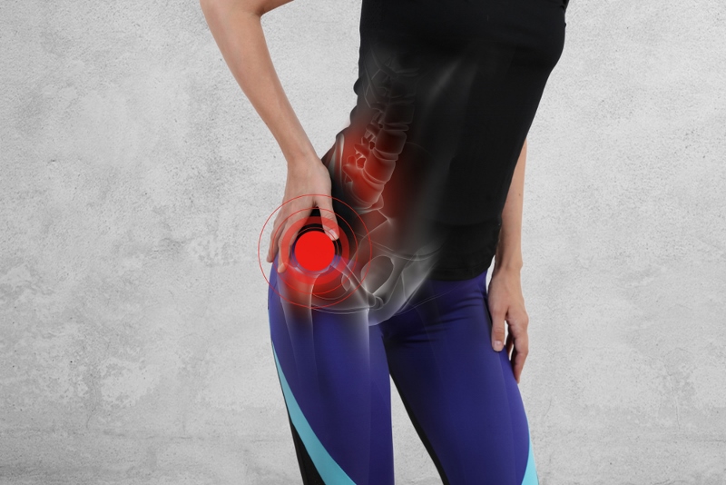
Hip Arthrosis
Hip osteoarthritis or coxarthrosis is a chronic inflammatory disease due to the wear and tear of the articular cartilage present in the hip joint.
LEARN MORE
SYMPTOMS
It usually manifests with pain in the groin.
CAUSES
Coxarthrosis is rarely due to a single cause; in general, the combination of several factors favors and leads to the generation of the articular cartilage of the femoral head and acetabulum and consequent thinning. The most relevant factors are: advanced age, trauma or fractures affecting the bone portions constituting the hip, joint or bone infections, congenital diseases of the hip and osteonecrosis.
DIAGNOSIS
Through a series of diagnostic imaging tests including hip X-rays and MRI of the hip.
THERAPY
Treatment of hip osteoarthritis depends on the symptoms and stage of the condition. In the presence of coxarthrosis in the first stage, conservative therapy is opted for; in the case of an advanced stage of coxarthrosis, surgical therapy (hip prosthesis) or regenerative medicine is chosen.
Conservative therapy is generally given by:
- Rest
- Drugs: NSAIDs, paracetamol, chondroprotectors (ex. Glucosamine), corticosteroids, hyaluronic acid, PRP guided by ultrasound.
- Physiotherapy
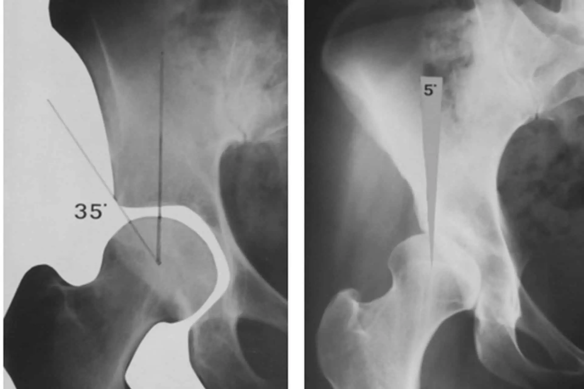
Hip Joint Instability
Hip joint instability is a condition in which the hip joint is excessively loose or prone to dislocation.
LEARN MORE
INJURY
The hip joint is very stable thanks to its structure. The femoral head is almost entirely inserted into the acetabulum and this establishes a firm union between the two bones, strengthened by the acetabular labrum.
The capsule-ligament structure and other ligaments exert a strong containment (stabilizing) action that keeps the femoral head in place. The muscles also act as dynamic stabilizers.
Hip instability is a rare disease and mainly affects sportsmen (golf, track and field, weightlifters, etc.). It can be difficult to diagnose and has varying degrees of severity.
CAUSES
Hip instability is determined by an excessive freedom of movement of the femoral head within the acetabulum.
The excessive movement is due to pathologies of the containment structures of the joint: for example, the laxity of the capsule-ligament structure -which can also be congenital- and capsule-ligament injuries caused by high-energy trauma such as fractures and dislocations.
Even lesions of the acetabular lip, more rarely of the tendons and muscles, can alter the biomechanics of the joint, leading to instability.
Hip instability can also be caused by morphological abnormalities, such as in hip developmental dysplasia.
When dysplasia is diagnosed at birth, it can be treated with excellent results, however when it is diagnosed in adulthood, the instability has had time to cause more or less serious damage.
In more extreme cases, migration of the femoral head beyond the acetabulum (inveterate hip dislocation) is observed.
TREATMENT
- Treatments vary according to the type of lesion, for example, capsulo-ligamentous and lip lesions can be treated with arthroscopy.
- Physiotherapy is essential to rebalance the functionality of the limb and to strengthen and tone the muscles that help stabilize the joint.
- Adequate musculature is also very important in preventing hip instability, especially for athletes.
Acetabular Labrum Injury
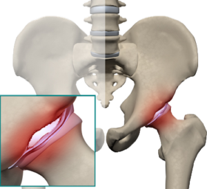
Acetabular labrum rupture is damage to the cartilaginous ring surrounding the hip socket.
LEARN MORE
CAUSES
The abnormal shape of the acetabulum and/or femoral head is one of the most frequent causes of the problem. One of the most common anatomical abnormalities that predispose to acetabular labrum injuries is Femoro Acetabolar Impingement.
SYMPTOMS
The main symptoms are:
- Pain in the front of the hip accompanied by a clicking or locking sensation in the hip.
- Pain can be constant and can limit all sporting and recreational activities.
DIAGNOSIS
Your doctor will run a few tests, with the most common being the sign of impingement. This test is performed by bending the hip to 90 degrees (flexion), rotating the hip inward (internal rotation), and bringing the thigh toward the other hip (adduction). Hip x-rays and an MRI of the hip are always helpful.
TREATMENT
- Physiotherapy;
- Surgical treatment
Snapping Hip Syndrome
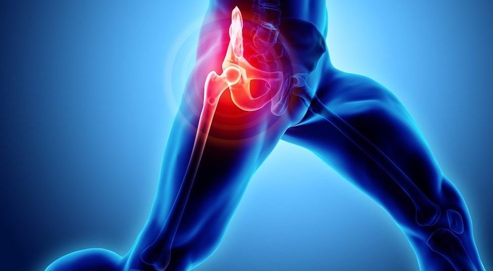
The snapping hip or snapping hip syndrome occurs in particular situations of movement: running or carrying out movements that involve the hip.
LEARN MORE
SYMPTOMS
Snapping hip syndrome generally does not manifest itself through pain but through clicking or crunching noises. What the patient feels is a sense of discomfort during movements: he may also feel joint or bone pain concerning the trochanteric part, the gluteal region, and the inguinal region.
CAUSES
They are not well known but probably some could be:
- Connective tissue weakness;
- Different length of the lower limbs;
- Previous hip injuries;
DIAGNOSIS
Snapping hip syndrome is diagnosed by investigating previous injuries or current symptoms, followed by a physical examination by a doctor. The purpose of this is to find the cause of snapping by turning the hip more than once in various directions. It is always good to deepen the diagnosis by carrying out tests such as radiography and magnetic resonance imaging
TREATMENT
Among the more conservative treatments we find:
- Use of glucocorticoids or anesthetics;
- Ice on the affected area;
- Perform stretching before and after sports activities.
Avascular Necrosis of the Femoral Head
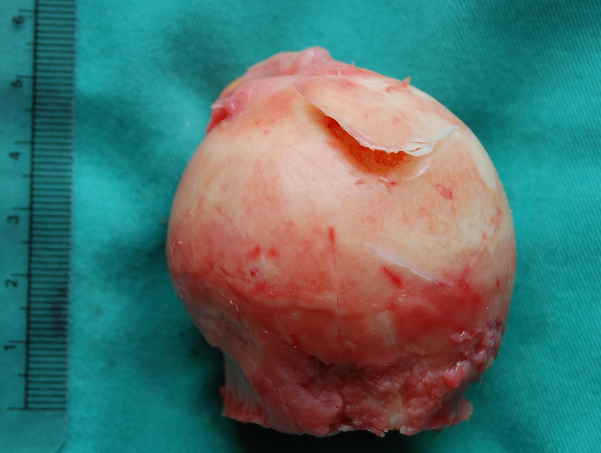
Avascular necrosis of the femoral head is the loss of blood supply to the hip bone, leading to bone death and potential collapse of the femoral head.
LEARN MORE
DIAGNOSIS
Have you been diagnosed with suspected avascular necrosis of the femoral head?
What is it about?
Necrosis of the femoral head is the result of a vascularization defect of the femoral head and consequent resorption of the bone tissue in a delimited area, inducing a degeneration of the entire joint.
It is a more frequent pathology in males between the ages of thirty and sixty and can be bilateral.
CAUSES
The causes can be multiple, both of a traumatic or non-traumatic nature.
In the case of a trauma, we can have a vascular lesion and a decrease in the perfusion of the femoral head. In non-traumatic conditions, it is often difficult to understand the causes. Often, the etiopathogenesis of osteonecrosis is determined by concomitant factors: drug intake (eg cortisone for long periods), alcohol, diabetes, metabolic diseases, coagulation disorders, etc.
SYMPTOMS
The patient begins to experience pain in the hip, especially after exertion and in some movements (eg putting on stockings) with consequent lameness. Pain is usually the symptom of onset and has a sudden onset. It can be referred to the groin, radiated to the anterior or anteromedial aspect of the thigh and, more rarely, to the buttock. Pain is often present, even at rest, but loading and walking aggravate it significantly.
DIAGNOSIS
Traditional radiographs may show modest density changes in the bone texture or may show changes in the morphology of the deformed femoral head. The first radiographic findings of osteonecrosis can be evident even after about 3 months. The CT scan can help to define the extent of the lesion and the degree of bone collapse in the most advanced states, but the MRI is the diagnostic method of choice as it allows to discover the lesions early on even before they are visible in traditional radiographs.
TREATMENT
Initially, treatment consists of limb unloading, osteoporosis-like drug therapy, and physical therapies such as magneto therapy and hyperbaric oxygen therapy. Growth factors are successfully used in the initial stages of the disease, however it is a difficult challenge since recovery is not always achieved; moreover, in the earliest stages of the lesion, the patient is often asymptomatic and the disorders appear when the treatment options are only surgical and in young patients with high functional demands, prosthetic hip replacement is at greater risk of wear and failure. In consideration of the early age in which the disease often manifests itself and, consequently, of the high functional demands, the importance of “biological” therapeutic options emerges that allow to spare the joint, avoiding the collapse of the femoral head.
The research perspectives on which we have been focusing on for some years are aimed at the study of the pathophysiology of the disease and the use of autologous growth factors and mesenchymal cells that are proving to be fundamental in promoting the processes of reparative osteogenesis.
RECOVERY
After the surgical treatment, it is necessary to limit the load on the hip and follow a careful rehabilitation protocol.
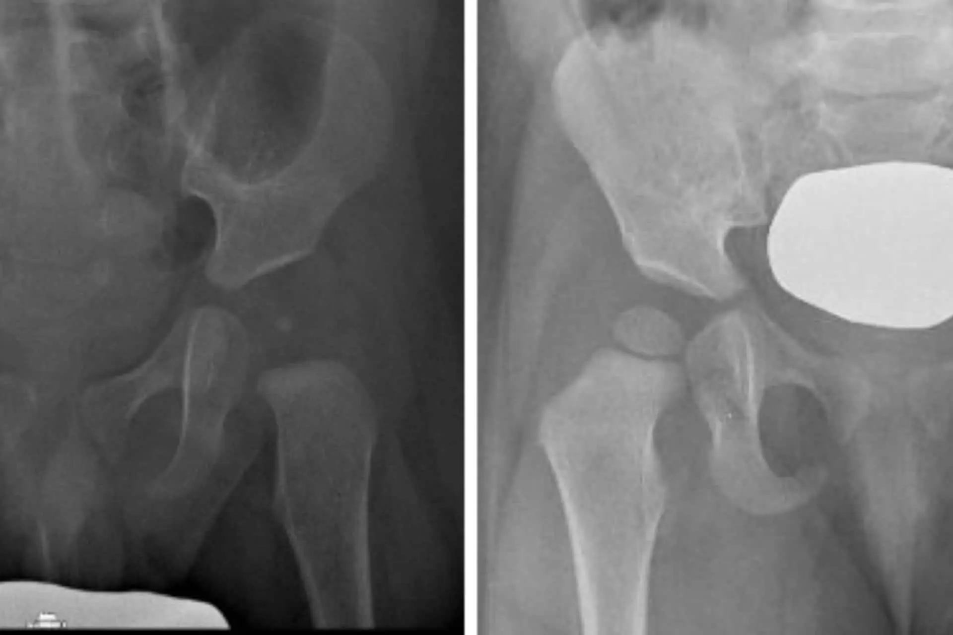
Developmental Dysplasia of the Hip
Developmental dysplasia of the hip is the most common among childhood hip diseases, mainly affecting females and those born breech.
LEARN MORE
Developmental dysplasia of the hip is the most common among childhood hip diseases, mainly affecting females and those born breech.
The pathology has different degrees of severity from birth: the hip may be unstable, therefore it tends to dislocate, or may already be dislocated, due to capsule-ligament laxity and/or acetabular abnormalities that prevent the femoral head from articulating correctly with the acetabulum.