


Painful Syndromes
Discover less common pathologies that our doctors can help you treat
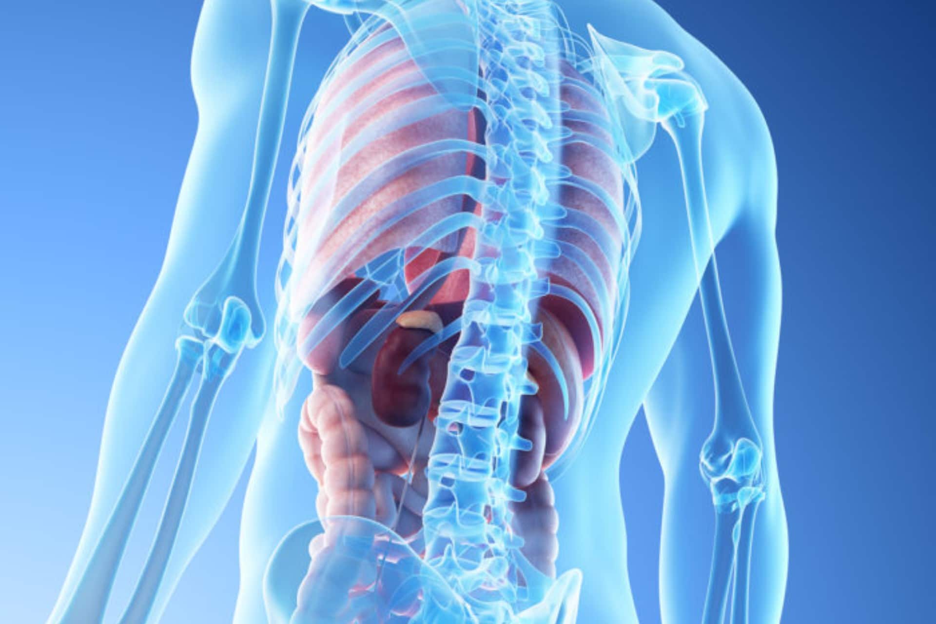
Cervical Thoracic Interstitial Bursitis
The pain is localized in the interstitial region between C7 and Th1 and does not radiate. It is constant, aching. The patient can try to reduce pain by adopting a kyphosis pose with the neck extended forward.
Learn More
The pain is localized in the interstitial region between C7 and Th1 and does not radiate. It is constant, aching. The patient can try to reduce pain by adopting a kyphosis pose with the neck extended forward.
Pain with cervicothoracic interstitial bursitis often decreases during movement and increases at rest.
The examination reveals pain with deep palpation of the C7-Th1 region, often with a reflex spasm of the paravertebral muscles. There is always a limitation of movement and an increase in pain during flexion in the cervical and upper chest areas.
There is no specific method for detecting cervico-thoracic interstitial bursitis.
The examination, first of all, is aimed at revealing the hidden pathology or other diseases that can mimic cervico-thoracic interstitial bursitis.
X-ray examination can reveal any changes in the bones of the cervical region, including arthritis, fracture, congenital pathology (Arnold-Chiari malformation) and a tumor.
All patients with recent onset of cervico-thoracic interstitial bursitis should undergo MRI of the cervical spine and brain if symptoms of occipital and significant headache are present.
To rule out hidden inflammatory arthritis, infection and cancer, a laboratory screening must be performed, including a general blood test, an ESR, an antinuclear antibody test and a biochemical blood test.

Women and ACL Injuries
For more than a decade, researchers have debated various reasons as to why anterior cruciate ligament (ACL) injuries affect women more than men, considering both anatomical and hormonal differences between the two sexes.
Learn More
For more than a decade, researchers have debated various reasons as to why anterior cruciate ligament (ACL) injuries affect women more than men, considering both anatomical and hormonal differences between the two sexes.
Recent studies show that female athletes who play certain sports such as football or basketball are 3-4 times more prone to ACL injury than male athletes.
Most of these injuries occur in women between the ages of 15 and 25.
At the June 1999 conference sponsored by the American Academy of Orthopedic Surgeons, researchers reported the following factors as an explanation for the increased incidence of ACL injury in female athletes.
- Biomechanical factors. Experts report that women tend to pay more attention to their quadriceps muscles than men, believing this to be the main reason for the increased risk of ACL injury. All researchers agree that women should learn to use their tendons more often. The experts also concluded that women tend to place their foot on the sole rather than on the toes and this may contribute to the increase in the incidence of injuries.
- Hormonal influences Experts argue that these should not change the level of activity and it is not necessary to abstain from sports during the menstrual cycle. They also argue that female hormones do not increase the chances of suffering an ACL injury, but suggest that further investigation is warranted.
- Environmental factors. The use of braces does not prevent ACL injuries. Furthermore, the use of specific footwear can on the one hand increase performance because they guarantee a good grip on the ground, but at the same time increase the risk of injury.
- Anatomical factors. There is insufficient data to support the theory that ACL size correlates with injury risk.
Researchers agree that training programs that teach how to land the foot properly and basic injury prevention techniques should be adopted to help female athletes.
The two main sports considered to be at greatest risk of ACL injury in women are basketball and football.

Eagle Syndrome
Eagle’s syndrome or styloid syndrome is a disorder of the ligaments that connect the skull to the hyoid bone in the neck, which following calcification cause painful symptoms in the mouth, head, neck, throat and forehead. In particular, it is caused by the lengthening of the styloid process, a small bone at the base of the skull and / or by calcification of the ligament.
Learn More
WHAT IS EAGLE SYNDROME
Eagle’s syndrome or styloid syndrome is a disorder of the ligaments that connect the skull to the hyoid bone in the neck, which following calcification cause painful symptoms in the mouth, head, neck, throat and forehead. In particular, it is caused by the lengthening of the styloid process, a small bone at the base of the skull and / or by calcification of the ligament.
The condition typically occurs after an injury or trauma to the neck or pharynx or after an operation involving the neck sector, such as tonsillectomy.
SYMPTOMS
Manifestations of Eagle syndrome include:
- A nagging or blunt pain in the throat, head, neck and face
- Pain that persists for long durations over days, weeks or even months
- Pain in the ear on the same side as pain in the head, neck, face
- Sensation of something stuck in the throat
- Pain in swallowing or oodynophagy
- Hoarseness of the voice or dysphonia
- Excessive salivation or drooling
- Other symptoms may include altered voice, cough, dizziness, migraine, occipital neuralgia, pain in the teeth and jaw, and sinus or bloodshot eyes.
DIAGNOSIS
Diagnosis is commonly made using x-ray, CT, or MRI scans which can reveal calcification of the stylohyoid ligament.
TREATMENT
The treatment primarily involves the use of a personalized biological therapy, PRP (Platelet-Rich Plasma), which may help relieve pain.
In most patients, this approach has proven beneficial, with a low risk of recurrence. In severe recurrent cases, surgical intervention may become necessary.
At our center, we perform local injections under ultrasound guidance, often providing significant clinical benefit.
Headache
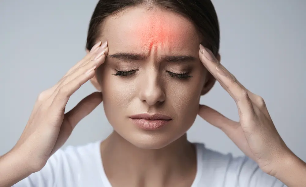
Headache is a nagging pain in any part of the head including the upper neck and face; headaches are one of the most common reasons for medical consultation as they interfere with the ability to work and perform daily activities. Some people suffer from frequent headaches. Although headaches can be painful and stressful, they are rarely caused by a serious condition
Learn More
Headache is a nagging pain in any part of the head including the upper neck and face; headaches are one of the most common reasons for medical consultation as they interfere with the ability to work and perform daily activities. Some people suffer from frequent headaches. Although headaches can be painful and stressful, they are rarely caused by a serious condition. The
headaches can be divided into two types:
Primary headaches: not caused by another disorder
Secondary headaches: caused by another disorder
Primary headache disorders include migraine, cluster headache, and muscle tension headache.
Secondary headaches can result from disorders of the brain, eyes, nose, throat, sinuses, teeth, jaws, ears, or nape of the neck, or from a widespread (systemic) disorder throughout the body. Some secondary headaches are severe.
In particular those involving the brain, such as meningitis, a brain tumor or haemorrhage.
Fever can cause headaches, as can many infections that don’t necessarily involve the brain. Headaches are also common when you stop consuming caffeine or taking medications.
Pain relievers (analgesics) after using them for a long time (drug overuse headache).
Contrary to what most people think, eye strain and arterial hypertension (except for the extreme one) do not usually cause headaches.
When to seek immediate medical attention
Very sudden and severe headache, changes in sensation or vision, sudden weakness, loss of coordination, convulsions, difficulty in speaking or understanding speech, changes in
levels of consciousness, such as sleepiness or confusion (suggesting a brain disorder), fever and a stiff neck, which makes it painful or sometimes impossible to lower the chin towards the chest.
Exams
Most people do not need tests as they do not have a serious cause, particularly if they start at a young age, if they have not changed over time and if the results of the physical examination are normal.
However, if the doctor suspects a disease, magnetic resonance imaging (MRI) or computed tomography (CT) may be requested.
A spinal tap (lumbar puncture) is usually done if acute meningitis or encephalitis (brain infection) is suspected.

Headaches Related to Sexual Activity
Generally, sexually associated headaches comprise a group of benign disorders, but in rare cases, the patient may experience an acute subarachnoid hemorrhage during sexual activity, which can be misdiagnosed as a benign explosive headache. There is no gender predilection for sexually associated headaches, and their onset may be episodic or chronic. Rarely, more than one type of sexually associated headache occurs in the same patient.
Learn More
WHAT IS IT
Doctors have identified the following three general types of headache associated with sexual activity:
- Explosive type
- Opaque type
- Postural type
Generally, sexually associated headaches comprise a group of benign disorders, but in rare cases, the patient may experience an acute subarachnoid hemorrhage during sexual activity, which can be misdiagnosed as a benign explosive headache. There is no gender predilection for sexually associated headaches, and their onset may be episodic or chronic. Rarely, more than one type of sexually associated headache occurs in the same patient.
SINTOMI
Patients with this disorder present differently depending on the type of headache they are affected by:
Headache associated with explosive sexual activity
The explosive type of sexual headache is the most common type encountered in clinical practice. Patients usually fear they have had a stroke. It occurs suddenly, with an almost instantaneous onset that peaks shortly before or during orgasm. The intensity of the explosive type is severe. The pain is usually localized in the occipital region. The pain is usually bilateral. The pain usually remains intense for 10–15 minutes and then gradually subsides. Some patients experience residual headache pain for 2 days.
Muscle tension headache associated with sexual activity
This type of headache begins during the initial stages of sexual activity and has a muscle-tension nature, starting in the occipital region. The headache becomes holocranial as sexual activity progresses toward orgasm. It may peak at orgasm, but unlike the explosive type, this type of pain is “dull” and disappears quickly after orgasm.
Postural type of headache associated with sexual activity
The postural type of sexual headache is similar to the explosive type in that it occurs shortly before or during orgasm. Its rapid onset at the peak and its severe intensity are also similar to those of the explosive type. It differs from the explosive type in that the headache symptoms appear when the patient stands up, after sexual activity itself.
DIAGNOSIS
Brain magnetic resonance imaging (MRI) provides the best information about the cranial vault and its contents. More importantly, MRI helps identify hemorrhages associated with leaking intracranial aneurysms. In patients who cannot undergo MRI, such as those with pacemakers, computed tomography (CT) is a reasonable second choice.
If the diagnosis of sexual headache is questionable, screening tests should be performed, including a complete blood count, erythrocyte sedimentation rate (ESR), and blood chemistry analysis. Intraocular pressure should be measured if glaucoma is suspected.
TREATMENT
It is generally believed that avoiding sexual activity for a few weeks reduces the likelihood of triggering sexual headaches. If this avoidance technique fails or is impractical due to patient preference, the next step is to use propranolol. If propranolol is ineffective, indomethacin can be tried. Indomethacin should be used with caution.
Cough Headache
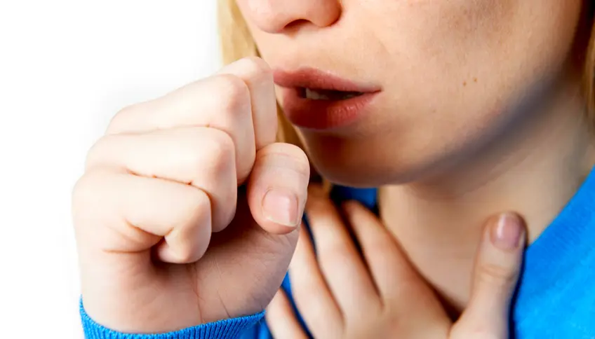
Cough headache is a term used to describe headaches triggered by coughing and other activities associated with a Valsalva maneuver, such as laughing, straining at stool, lifting, and bending the head toward the ground.
Learn More
WHAT IS IT
Cough headache is a term used to describe headaches triggered by coughing and other activities associated with a Valsalva maneuver, such as laughing, straining at stool, lifting, and bending the head toward the ground. Clinicians have identified the following two types of cough headache:
- Benign
- Symptomatic
Initially, both types of cough headache were thought to be related to sexual and exertional headaches, but they are now considered distinct clinical entities. A strong male predilection is seen for benign cough headache and no gender predilection for symptomatic cough headache.
SYMPTOMS
Patients suffering from cough headache present differently depending on the type of cough headache experienced. Each clinical presentation is discussed.
Benign Cough Headache
Benign cough headache is not associated with obvious neurological or musculoskeletal disease. More than 80% of patients with benign cough headache are males, in contradistinction to symptomatic cough headache, in which no gender predilection is seen. The onset of benign cough headache is abrupt, occurring immediately after coughing or other activities that cause a Valsalva maneuver. Although the intensity of pain is severe and peaks rapidly, it lasts only seconds to minutes. The character of the pain associated with benign cough headache is splitting or sharp, and the pain is located in the occipital region bilaterally and occasionally the vertex of the skull.
Symptomatic Cough Headache
Symptomatic cough headache is almost always associated with structural abnormalities of the cranium. Similar to benign cough headache, the onset of pain associated with symptomatic cough headache is abrupt, occurring immediately after coughing or other activities that cause a Valsalva maneuver. Although the intensity of pain is severe and peaks rapidly, it lasts only seconds to minutes. In contrast to benign cough headache, associated neurological symptoms may be present, including difficulty swallowing, faintness, and numbness in the face and upper extremities. These associated symptoms should be taken very seriously because they are indicative of increased intracranial pressure and herniation of the intracranial contents. The character of the pain associated with symptomatic cough headache is splitting or sharp, and pain is located in the occipital region bilaterally and occasionally the vertex of the skull.
DIAGNOSIS
Magnetic resonance imaging (MRI) of the brain provides the best information regarding the cranial vault and its contents. MRI is highly accurate and helps identify abnormalities that may put the patient at risk for neurological disasters secondary to intracranial and brainstem pathological conditions. In patients who cannot undergo MRI, such as patients with pacemakers, computed tomography (CT) is a reasonable second choice.
Screening laboratory tests consisting of complete blood cell count, erythrocyte sedimentation rate, and automated blood chemistry testing should be performed if the diagnosis of cough headache is in question. Intraocular pressure should be measured if glaucoma is suspected.
TREATMENT
Indomethacin is the treatment of choice for benign cough headache. Indomethacin must be used carefully.

Hypnic Headache
Also known as “wake-up headache,” hypnic headache is a term used to describe an uncommon headache syndrome characterized by the patient waking up at the same time every night. It always occurs during sleep. Hypnic headaches are short-lived and rarely last more than 15 minutes after the patient is awakened by the pain. Unlike cluster headaches, which also occur after sleep, patients with hypnic headaches show no autonomous signs or symptoms. Hypnic headaches are a disorder that mostly begin after age 50. They occur more commonly in women.
Learn More
DIAGNOSIS
Magnetic resonance imaging (MRI) of the brain provides the best information. Plain radiographs of the cervical spine can also be helpful in evaluating Arnold-Chiari malformations and should be included in the evaluation of all patients with hypnic headache.
If the diagnosis of hypnic headache is in doubt, screening laboratory tests should be performed, consisting of a complete blood count, erythrocyte sedimentation rate, and automated blood chemistry analysis.
Nighttime headaches that can be confused with hypnotic headaches:
- Cluster headache
- Headaches associated with sleep apnea
- Headache associated with nocturnal arterial hypertension
- Headache associated with increased intracranial pressure
- Analgesic rebound headache.
TREATMENT
Indomethacin and lithium carbonate are the preferred treatments for hypnotic headache.
Indomethacin should be used with caution, especially in patients with peptic ulcer disease or impaired renal function. The therapeutic window for lithium carbonate is small, so this drug should be used with caution. Unlike cluster headache, inhaled oxygen has been ineffective in aborting hypnic headache attacks.

Infraspinatus Tendinitis
Infraspinatus tendinitis can manifest as an acute or chronic painful condition of the shoulder. It usually occurs in a younger group of patients following overuse or misuse of the shoulder joint. Contributing factors include activities that require repetitive abduction and lateral rotation of the humerus, such as installing brake pads while working on an assembly line. Vigorous use of exercise equipment has also been implicated.
Learn More
SYMPTOMS
The pain of infraspinatus tendinitis is constant, severe, and localized to the deltoid area. Significant sleep disturbances are often reported. Chronic infraspinatus tendinitis tends to occur in older patients and develops more gradually or insidiously, without a single specific antecedent trauma. The pain may be associated with a gradual loss of range of motion in the affected shoulder. The patient often wakes at night when rolling over on the affected shoulder.
DIAGNOSIS
Plain radiographs are indicated for all patients with shoulder pain. Depending on the patient’s clinical presentation, additional tests may be indicated, including a complete blood count, erythrocyte sedimentation rate, and antinuclear antibody test. Magnetic resonance imaging (MRI) of the shoulder is indicated if a rotator cuff tear is suspected and to confirm the diagnosis of infraspinatus tendinitis.
TREATMENT
Initial treatment should include a combination of nonsteroidal anti-inflammatory drugs (NSAIDs) or cyclooxygenase-2 (COX-2) inhibitors and physical therapy. Local heat and cold application may also be helpful. Vigorous exercise should be avoided. Bupivacaine and methylprednisolone injections may be considered if physical therapy fails to achieve the desired results.

Post-dural Headache
When the dura mater is intentionally or accidentally perforated, headache may occur. The clinical presentation of postdural puncture headache is classic and confirms the diagnosis. The symptoms and rare physical findings associated with postdural puncture headache are due to low cerebrospinal fluid pressure caused by the continuous leakage of spinal fluid from the subarachnoid space.
Learn More
SYMPTOMS
Symptoms of postdural puncture headache begin almost immediately after the patient moves from a horizontal to an upright position. The intensity peaks within 1 to 2 minutes and subsides within a few minutes of the patient returning to a horizontal position. The headache has a throbbing character and is severe in intensity, increasing the longer the patient remains upright. The headache is almost always bilateral and localizes to the frontal, temporal, and occipital regions. Nausea, vomiting, and dizziness often accompany the headache, especially if the patient remains upright for prolonged periods. Visual disturbances may occur in cases of cranial nerve palsy.
DIAGNOSIS
Magnetic resonance imaging (MRI) with and without gadolinium is very accurate in confirming the diagnosis of postdural headache.
No additional tests are indicated for a patient who has undergone a dural puncture and subsequently develops a classic postural headache, unless infection or subarachnoid hemorrhage is suspected. In this case, a lumbar puncture, complete blood count, and erythrocyte sedimentation rate are indicated in an emergency.
TREATMENT
The cornerstone of postdural puncture headache treatment is the administration of autologous blood into the epidural space. This technique is known as epidural blood patching and is highly effective in treating postdural puncture headache. The patient must remain in a horizontal position for the next 12–24 hours. Approximately 10% of patients experience temporary relief and then a recurrence of symptoms when they stand up.
If the patient has suffered from significant nausea and vomiting, antiemetics may help.
Parsonage Turner Syndrome
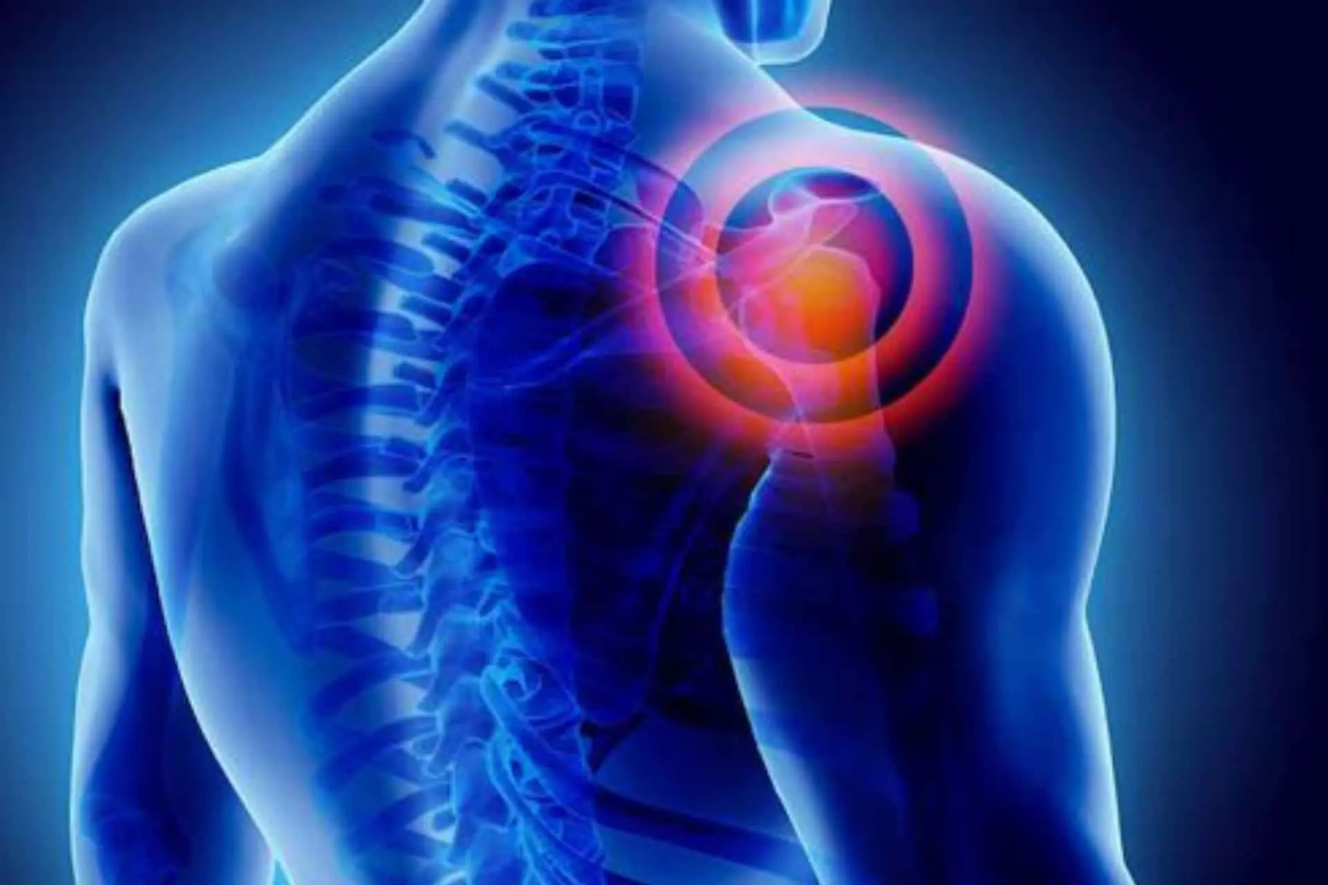
This syndrome affects the shoulder and upper limbs and takes its name from the person who first described it in 1948.
The pain is acute and burning, such as to prevent even sleep, precedes the muscle weakness that appears a few days later.
A viral etiology is assumed but some hypothesize an immunological disease
Learn More
SYMPTOMS
The pathology manifests itself with sudden and acute pain in the shoulder radiating to the upper limb at the level of the deltoid, supra and infraspinatus, biceps and triceps.
DIAGNOSIS
It is difficult because it is often confused with a neuropathy of the brachial plexus or with the thoracic outlet syndrome; it is also necessary to consider a possible compressive effect of the plexus of tumor origin (Pancoast Syndrome) in addition to trauma to the plexus.
The electromyographic examination can be of great use if positive due to plexus suffering, associated with MRI of the cervical spine and brachial plexus as well as blood chemistry tests (antinuclear antibodies).
TREATMENT
Pharmacological therapy involves the use of Gabapentinoids, Carbamazepine, Baclofen and other muscle relaxants.
Blockade of the brachial plexus with anesthetics and corticosteroids can represent the only solution for uncontrolled pain.
Physiotherapy treatments and physical therapies.
Recovery is slow and requires supportive therapies for the patient who can develop mental health issues due to severe pain.

Photosensitivity from drugs
Photosensitivity is a skin reaction that becomes particularly sensitive to damage induced by sun exposure.
It manifests itself with an important skin reaction: redness, itching and burns with formation of blisters after exposure to sunlight.
The most common phototoxic reactions tend to occur a few hours after exposure to the sun.
Learn More
CAUSE
Many chemicals are able to cause photoallergic reactions, from drugs used for various diseases (antibiotics, sulfonamides, antifungals, cardiovascular and anti-arrhythmic, anti-inflammatory drugs, etc.) to substances that can be applied to the skin e.g. after shave lotions, sun creams, etc.
This type of photosensitivity can occur after exposure to the sun, even after terminating therapy with the drug, and can sometimes spread to areas of the skin surface that have not been directly exposed to the sun.
SYMPTOMS
This type of photosensitivity can occur after exposure to the sun, even after terminating therapy with the drug, and can sometimes spread to areas of the skin surface that have not been directly exposed to the sun.
These skin conditions have the characteristics of urticaria, with redness, peeling and sometimes blisters and spots.
Individuals with phototoxic reactions may initially complain of a burning sensation and in the following hours redness appears in areas exposed to the sun, such as the forehead, nose, hands, arms, legs and feet; the extent of skin damage can range from a slight redness to the formation of blisters and bubbles.
Some medical conditions are aggravated by sun exposure: systemic lupus erythematosus (SLE) often causes a rash on the face and can be very sensitive to sun exposure.
Porphyria is a hereditary disease characterized by a cutaneous manifestation, which can worsen following the stimulus represented by sunlight, with rashes and blisters.
Vitiligo is a relatively common disease that occurs with depigmented skin spots, which lack melanin and are extremely sensitive to UV rays.
TREATMENT
If possible, discontinue the photosensitizing drug immediately.
These phenomena are often reversible and resolve spontaneously.
Those who are photosensitive or who take drugs that can cause photosensitivity should minimize exposure to the sun and use sunscreens and protective clothing.
Local dressings can relieve the symptoms of photodermatosis: topical application of creams with cortisone can be useful in the treatment of redness, while antihistamines can reduce itching. In severe cases, a short therapy (10-14 days) of oral corticosteroids can be practiced.
People with other concomitant dermatological conditions should see a doctor right away.
Pubalgia
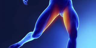
Pubalgia is a syndrome characterized by pain in the groin and pubic area, sometimes radiating to the inside of the thighs.
Learn More
WHAT IS IT
Pubalgia is a syndrome characterized by pain in the groin and pubic area, sometimes radiating to the inside of the thighs.
Its etiology is wide but they are generally distinguished:
An INSERTIONAL TENDINOPATHY caused by repeated microtrauma to the adductor muscles of the thigh and abdominal muscles.
A pathology of the ABDOMINAL RECTUM often caused by the act of kicking during which there is a strong tension in the abdominal muscles. This tension sometimes creates a fissure in the superficial fascia with stretching and compression of the perforating nerve.
PUBIC SYMPHYSIS SYNDROME: caused by microtrauma of the adductor muscles, a situation that arises especially in the age of development.
SYMPTOMS
Si può presentare con un dolore soprattutto in alcuni movimenti specifici dell’attività fisica.It can present with pain especially in some specific movements of physical activity.
In the most severe cases, the pain is continuous and worsens with sudden movements.
n clinical examination, the adductor muscles appear to be contracted and the pressure at the level of the pubis is more or less painful.
DIAGNOSIS
Clinical history and clinical examination are very important. Tests such as ultrasound and magnetic resonance are generally used to exclude other pathologies (eg. A hernia). An accurate postural examination must exclude positional defects or pathologies of the lower limbs that cause imbalance of the pelvis.
TREATMENT
Early diagnosis allows for effective treatment. In groin pain it is necessary to be able to understand what is the triggering cause and try to eliminate it. Various medical, physical or kinesiological therapies are adopted. Among the physical treatments, laser, Tekar therapy and shock waves deserve mention. Among medical therapies, infiltrations with hyaluronic acid and more recently with PRP (Platelet Rich Plasma) are often used.
Sports rest is recommended.
From the kinesiological point of view, exercises to lengthen the adductor muscles and the posterior chains with Mezieres-type exercises are recommended. Also coordination and reprogramming of the movement pattern in the gym.
Paget's Disease
Paget’s disease is a metabolic bone disease, also known as osteitis deformans or Paget’s disease of bone.
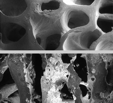
The disease involves an alteration of bone remodeling, which occurs at an exaggerated and disordered rate, undermining the structural integrity of the skeleton and predisposing to fractures and bone malformations.
Learn More
SYMPTOMS
Paget’s disease can present insidiously, asymptomaticly, or with mild symptoms. In many cases, the diagnosis is made accidentally or only after complications develop. The most common complaint is bone pain. The disease can affect individual bones or spread to multiple areas of the body. In some cases, skin temperature may increase in areas where the disease is active.
Common symptoms of Paget’s disease may include: bone pain and joint stiffness, numbness, tingling, and weakness;
Bone deformities: enlargement of the affected segments and other visible deformities, such as those affecting the skull (frontal bossing, enlarged jaw or increased head size).
DIAGNOSIS
Diagnosis of Paget’s disease can be made through X-rays, which are the first-line investigation. They provide radiological evidence of the affected skeletal segment (areas of bone resorption, bone enlargement, and deformity) and are the most important diagnostic tool for identifying any complications. X-rays can be total body, pelvic, or lumbar spine, depending on the location of the disease. CT scans can aid in differential diagnosis (arthrosis, osteoporosis, osteomalacia, and bone metastases) and are useful for supplementing the information acquired through X-rays and monitoring the progression of the disease.
Bone scans are useful for determining the extent and activity of the disease. Their specificity is poor, but they are more sensitive than radiology in assessing the skeletal extent of Paget’s disease.
Blood and urine tests with higher than normal values of alkaline phosphatase in combination with unchanged parameters of calcium, phosphate and aminotransferases may indicate Paget’s disease of bone.
A bone biopsy is a rare investigation, especially if malignant progression is suspected. Pagetic bone is histologically characterized by a mosaic appearance.
TREATMENT
Currently, there is no definitive cure for Paget’s disease of bone , but some medications can help control the disease, relieve symptoms, and slow (or prevent) the onset of complications.
Choosing the right therapeutic strategy for patients with Paget’s disease can be complex.
The goal of drug therapy is to relieve bone pain and help prevent disease progression. Currently, the most widely used drugs are bisphosphonates , powerful inhibitors of bone resorption, which help control the disease and reduce pain and other symptoms.
Any complications often require specific treatment, such as: therapy for painful manifestations, anti-inflammatories to decompress the nerve roots, surgical interventions (nerve decompression, osteotomy or arthroplasty).